Respiratory System Anatomy and Physiology

Breathe life into your understanding with our guide on the respiratory system anatomy and physiology. Nursing students, immerse yourself in the intricate dance of inhalation and exhalation that fuels every living moment.

Table of Contents
Functions of the respiratory system, main bronchi, the respiratory membrane, respiration, mechanics of breathing, respiratory volumes and capacities, respiratory sounds, external respiration, gas transport, and internal respiration, control of respiration, age-related physiological changes in the respiratory system.
The functions of the respiratory system are:
- Oxygen supplier. The job of the respiratory system is to keep the body constantly supplied with oxygen.
- Elimination. Elimination of carbon dioxide.
- Gas exchange. The respiratory system organs oversee the gas exchanges that occur between the blood and the external environment.
- Passageway. Passageways that allow air to reach the lungs.
- Humidifier. Purify, humidify, and warm incoming air.
Anatomy of the Respiratory System
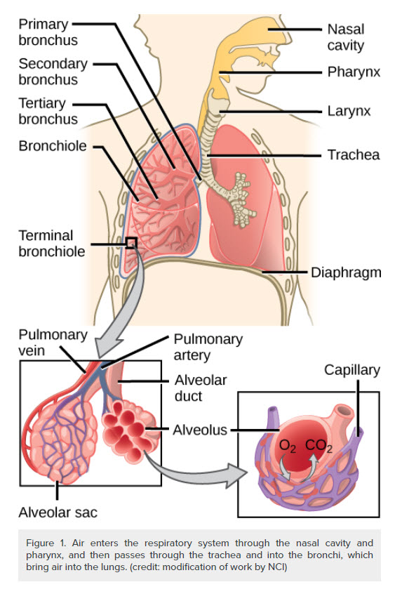
The organs of the respiratory system include the nose, pharynx, larynx, trachea, bronchi, and their smaller branches, and the lungs, which contain the alveoli.

The nose is the only externally visible part of the respiratory system.

- Nostrils. During breathing, air enters the nose by passing through the nostrils, or nares.
- Nasal cavity. The interior of the nose consists of the nasal cavity, divided by a midline nasal septum .
- Olfactory receptors. The olfactory receptors for the sense of smell are located in the mucosa in the slitlike superior part of the nasal cavity, just beneath the ethmoid bone.
- Respiratory mucosa. The rest of the mucosal lining, the nasal cavity called the respiratory mucosa, rests on a rich network of thin-walled veins that warms the air as it flows past.
- Mucus. In addition, the sticky mucus produced by the mucosa’s glands moistens the air and traps incoming bacteria and other foreign debris, and lysozyme enzymes in the mucus destroy bacteria chemically.
- Ciliated cells. The ciliated cells of the nasal mucosa create a gentle current that moves the sheet of contaminated mucus posteriorly toward the throat, where it is swallowed and digested by stomach juices.
- Conchae. The lateral walls of the nasal cavity are uneven owing to three mucosa-covered projections, or lobes called conchae, which greatly increase the surface area of the mucosa exposed to the air, and also increase the air turbulence in the nasal cavity.
- Palate. The nasal cavity is separated from the oral cavity below by a partition, the palate; anteriorly, where the palate is supported by bone, is the hard palate; the unsupported posterior part is the soft palate .
- Paranasal sinuses. The nasal cavity is surrounded by a ring of paranasal sinuses located in the frontal, sphenoid, ethmoid, and maxillary bones ; theses sinuses lighten the skull, and they act as a resonance chamber for speech.

- Size. The pharynx is a muscular passageway about 13 cm (5 inches) long that vaguely resembles a short length of red garden hose.
- Function. Commonly called the throat , the pharynx serves as a common passageway for food and air.
- Portions of the pharynx. Air enters the superior portion, the nasopharynx , from the nasal cavity and then descends through the oropharynx and laryngopharynx to enter the larynx below.
- Pharyngotympanic tube. The pharyngotympanic tubes, which drain the middle ear open into the nasopharynx.
- Pharyngeal tonsil. The pharyngeal tonsil, often called adenoid is located high in the nasopharynx.
- Palatine tonsils . The palatine tonsils are in the oropharynx at the end of the soft palate.
- Lingual tonsils . The lingual tonsils lie at the base of the tongue.
The larynx or voice box routes air and food into the proper channels and plays a role in speech.
- Structure. Located inferior to the pharynx, it is formed by eight rigid hyaline cartilages and a spoon-shaped flap of elastic cartilage, the epiglottis .
- Thyroid cartilage. The largest of the hyaline cartilages is the shield-shaped thyroid cartilage, which protrudes anteriorly and is commonly called Adam’s apple .
- Epiglottis. Sometimes referred to as the “guardian of the airways” , the epiglottis protects the superior opening of the larynx.
- Vocal folds. Part of the mucous membrane of the larynx forms a pair of folds, called the vocal folds, or true vocal cords , which vibrate with expelled air and allows us to speak.
- Glottis. The slitlike passageway between the vocal folds is the glottis.

- Length. Air entering the trachea or windpipe from the larynx travels down its length (10 to 12 cm or about 4 inches) to the level of the fifth thoracic vertebra , which is approximately midchest.
- Structure. The trachea is fairly rigid because its walls are reinforced with C-shaped rings of hyaline cartilage; the open parts of the rings abut the esophagus and allow it to expand anteriorly when we swallow a large piece of food, while the solid portions support the trachea walls and keep it patent, or open, in spite of the pressure changes that occur during breathing.
- Cilia. The trachea is lined with ciliated mucosa that beat continuously and in a direction opposite to that of the incoming air as they propel mucus, loaded with dust particles and other debris away from the lungs to the throat, where it can be swallowed or spat out.
- Structure. The right and left main (primary) bronchi are formed by the division of the trachea.
- Location. Each main bronchus runs obliquely before it plunges into the medial depression of the lung on its own side.
- Size. The right main bronchus is wider, shorter, and straighter than the left.

- Location. The lungs occupy the entire thoracic cavity except for the most central area, the mediastinum , which houses the heart, the great blood vessels, bronchi, esophagus, and other organs.
- Apex. The narrow, superior portion of each lung, the apex, is just deep into the clavicle.
- Base. The broad lung area resting on the diaphragm is the base.
- Division. Each lung is divided into lobes by fissures; the left lung has two lobes , and the right lung has three .
- Pleura. The surface of each lung is covered with a visceral serosa called the pulmonary , or visceral pleura, and the walls of the thoracic cavity are lined by the parietal pleura .
- Pleural fluid. The pleural membranes produce pleural fluid, a slippery serous secretion that allows the lungs to glide easily over the thorax wall during breathing movements and causes the two pleural layers to cling together.
- Pleural space. The lungs are held tightly to the thorax wall, and the pleural space is more of a potential space than an actual one.
- Bronchioles . The smallest of the conducting passageways are the bronchioles.
- Alveoli. The terminal bronchioles lead to the respiratory zone structures, even smaller conduits that eventually terminate in alveoli or air sacs.
- Respiratory zone. The respiratory zone, which includes the respiratory bronchioles, alveolar ducts, alveolar sacs, and alveoli, is the only site of gas exchange .
- Conducting zone structures. All other respiratory passages are conducting zone structures that serve as conduits to and from the respiratory zone.
- Stroma. The balance of the lung tissue, its stroma, is mainly elastic connective tissue that allows the lungs to recoil passively as we exhale.
- Wall structure. The walls of the alveoli are composed largely of a single, thin layer of squamous epithelial cells.
- Alveolar pores. Alveolar pores connect neighboring air sacs and provide alternative routes for air to reach alveoli whose feeder bronchioles have been clogged by mucus or otherwise blocked.
- Respiratory membrane. Together, the alveolar and capillary walls, their fused basement membranes, and occasional elastic fibers construct the respiratory membrane (air-blood barrier), which has gas (air) flowing past on one side and blood flowing past on the other.
- Alveolar macrophages. Remarkably efficient alveolar macrophages sometimes called “dust cells” , wander in and out of the alveoli picking up bacteria, carbon particles, and other debris.
- Cuboidal cells. Also scattered amid the epithelial cells that form most of the alveolar walls are chunky cuboidal cells, which produce a lipid (fat) molecule called surfactant , which coats the gas-exposed alveolar surfaces and is very important in lung function.
Physiology of the Respiratory System
The major function of the respiratory system is to supply the body with oxygen and to dispose of carbon dioxide. To do this, at least four distinct events, collectively called respiration, must occur.
- Pulmonary ventilation . Air must move into and out of the lungs so that gasses in the air sacs are continuously refreshed, and this process is commonly called breathing.
- External respiration. Gas exchange between the pulmonary blood and alveoli must take place.
- Respiratory gas transport. Oxygen and carbon dioxide must be transported to and from the lungs and tissue cells of the body via the bloodstream.
- Internal respiration. At systemic capillaries, gas exchanges must be made between the blood and tissue cells.
- Rule. Volume changes lead to pressure changes, which lead to the flow of gasses to equalize pressure.
- Inspiration. Air is flowing into the lungs; the chest is expanded laterally, the rib cage is elevated, and the diaphragm is depressed and flattened; lungs are stretched to the larger thoracic volume, causing the intrapulmonary pressure to fall and air to flow into the lungs.
- Expiration. Air is leaving the lungs; the chest is depressed and the lateral dimension is reduced, the rib cage is descended, and the diaphragm is elevated and dome-shaped; lungs recoil to a smaller volume, intrapulmonary pressure rises, and air flows out of the lung.
- Intrapulmonary volume. Intrapulmonary volume is the volume within the lungs.
- Intrapleural pressure. The normal pressure within the pleural space, the intrapleural pressure, is always negative, and this is the major factor preventing the collapse of the lungs.
- Nonrespiratory air movements. Nonrespiratory movements are a result of reflex activity, but some may be produced voluntarily such as coughing , sneezing, crying, laughing, hiccups, and yawning.

- Tidal volume. Normal quiet breathing moves approximately 500 ml of air into and out of the lungs with each breath.
- Inspiratory reserve volume. The amount of air that can be taken in forcibly over the tidal volume is the inspiratory reserve volume, which is normally between 2100 ml to 3200 ml.
- Expiratory reserve volume. The amount of air that can be forcibly exhaled after a tidal expiration, the expiratory reserve volume, is approximately 1200 ml.
- Residual volume. Even after the most strenuous expiration, about 1200 ml of air still remains in the lungs and it cannot be voluntarily expelled; this is called residual volume, and it is important because it allows gas exchange to go on continuously even between breaths and helps to keep the alveoli inflated.
- Vital capacity. The total amount of exchangeable air is typically around 4800 ml in healthy young men, and this respiratory capacity is the vital capacity, which is the sum of the tidal volume, inspiratory reserve volume, and expiratory reserve volume.
- Dead space volume. Much of the air that enters the respiratory tract remains in the conducting zone passageways and never reaches the alveoli; this is called the dead space volume and during a normal tidal breath, it amounts to about 150 ml.
- Functional volume. The functional volume, which is the air that actually reaches the respiratory zone and contributes to gas exchange, is about 350 ml.
- Spirometer. Respiratory capacities are measured with a spirometer, wherein as a person breathes, the volumes of air exhaled can be read on an indicator, which shows the changes in air volume inside the apparatus.
- Bronchial sounds. Bronchial sounds are produced by air rushing through the large respiratory passageways (trachea and bronchi).
- Vesicular breathing sounds. Vesicular breathing sounds occur as air fills the alveoli, and they are soft and resemble a muffled breeze.
- External respiration. External respiration or pulmonary gas exchange involves oxygen being loaded and carbon dioxide being unloaded from the blood.
- Internal respiration. In internal respiration or systemic capillary gas exchange, oxygen is unloaded and carbon dioxide is loaded into the blood.
- Gas transport. Oxygen is transported in the blood in two ways: most attaches to hemoglobin molecules inside the RBCs to form oxyhemoglobin, or a very small amount of oxygen is carried dissolved in the plasma; while carbon dioxide is transported in plasma as bicarbonate ion, or a smaller amount (between 20 to 30 percent of the transported carbon dioxide) is carried inside the RBCs bound to hemoglobin.
Neural Regulation
- Phrenic and intercostal nerves. These two nerves regulate the activity of the respiratory muscles, the diaphragm, and external intercostals.
- Medulla and pons. Neural centers that control respiratory rhythm and depth are located mainly in the medulla and pons; the medulla, which sets the basic rhythm of breathing, contains a pacemaker , or self-exciting inspiratory center, and an expiratory center that inhibits the pacemaker in a rhythmic way; pons centers appear to smooth out the basic rhythm of inspiration and expiration set by the medulla.
- Eupnea. The normal respiratory rate is referred to as eupnea, and it is maintained at a rate of 12 to 15 respirations/minute .
- Hyperpnea. During exercise, we breathe more vigorously and deeply because the brain centers send more impulses to the respiratory muscles, and this respiratory pattern is called hyperpnea.
Non-neural Factors Influencing Respiratory Rate and Depth
- Physical factors. Although the medulla’s respiratory centers set the basic rhythm of breathing, there is no question that physical factors such as talking, coughing, and exercising can modify both the rate and depth of breathing, as well as an increased body temperature, which increases the rate of breathing.
- Volition (conscious control). Voluntary control of breathing is limited, and the respiratory centers will simply ignore messages from the cortex (our wishes) when the oxygen supply in the blood is getting low or blood pH is falling .
- Emotional factors. Emotional factors also modify the rate and depth of breathing through reflexes initiated by emotional stimuli acting through centers in the hypothalamus .
- Chemical factors. The most important factors that modify respiratory rate and depth are chemical- the levels of carbon dioxide and oxygen in the blood; increased levels of carbon dioxide and decreased blood pH are the most important stimuli leading to an increase in the rate and depth of breathing, while a decrease in oxygen levels become important stimuli when the levels are dangerously low.
- Hyperventilation. Hyperventilation blows off more carbon dioxide and decreases the amount of carbonic acid, which returns blood pH to the normal range when carbon dioxide or other sources of acids begin to accumulate in the blood.
- Hypoventilation. Hypoventilation or extremely slow or shallow breathing allows carbon dioxide to accumulate in the blood and brings blood pH back into normal range when blood starts to become slightly alkaline.
Respiratory efficiency is reduced with age. They are unable to compensate for increased oxygen need and are significantly increasing the amount of air inspired. Therefore, difficulty in breathing is usually common especially during activities. Expiratory muscles become weaker so their cough efficiency is reduced and the amount of air left in the lungs is increased. Health promotion teaching can include smoking cessation, preventing respiratory infections through handwashing , and ensuring up to date influenza and pneumonia vaccinations.
Craving more insights? Dive into these related materials to enhance your study journey!
- Anatomy and Physiology Nursing Test Banks . This nursing test bank includes questions about Anatomy and Physiology and its related concepts such as: structure and functions of the human body, nursing care management of patients with conditions related to the different body systems.
10 thoughts on “Respiratory System Anatomy and Physiology”
Hello, My name is Sharon and I think I just struck gold! This web site is just what I need to study, learn, and understand the Human Body. Thank you so much.
Thank you so much. you just made my day. The information was comprehensive and comes as an easy resource. Violet.
Thanks very much, I was satisfied with the answers
Thanks very much. I am satisfied with the notes
Thank you so much the notes are perfect and useful to my studies
Very helpful and easy to understand. Thank you so much.
It is fantastic and very helpfull and thanks a lot
This is very helpful, thank you.
You’re welcome! Happy to hear that you found the respiratory system anatomy and physiology material helpful. If there’s anything more you’d like to dive into or any questions you have, feel free to reach out. Always here to help with your learning journey.
Thank you so much
Leave a Comment Cancel reply
Want to create or adapt books like this? Learn more about how Pressbooks supports open publishing practices.
Lab 6: Respiratory System
Measurable outcomes.
- Understand and identify the anatomical structures of the respiratory system on available models.
- Deduce the pathway of air through the respiratory system.
- Determine the pathway of pulmonary circulation.
- Identify the various muscles involved in respiration.
- Recognize the hallmarks of lung histology.
- Demonstrate an adequate understand of the material in this section.
The respiratory system is responsible for the gas exchange of oxygen and carbon dioxide. The main specialized organs of this process are the lungs which house clusters of sac-like structures known as alveoli . There are from 480 to 790 million alveoli which increase the efficiency of gas exchange by increasing surface area to around 118m 2 in men and 91m 2 in women. The respiratory system consists of the nasal cavity, pharynx, larynx, trachea, lungs, bronchi, bronchioles , and alveoli , along with their accessory structures. These structures are divided into the upper and lower respiratory systems , with the lower portion beginning at the larynx. The primary function of this system is to exchange oxygen and carbon dioxide between the body and the environment. Functionally, the respiratory system can be divided into the conducting zone , terminating at the terminal bronchioles; then air flows into the respiratory zone, where the actual gas exchange occurs.
Though we view each system individually in this lab, it is important to keep in mind that all organ systems overlap and work together in such a way that scientist are constantly discovering new connections. One such example is the nose . Not only is it the primary entrance and exit for respiration, but it also contains the olfactory epithelium, the primary structure of one of the special senses, olfaction. Likewise, the pharynx is a structure shared by both the respiratory and digestion systems.
Although both lungs functionally participate in respiration, they differ physically in various ways. The right lung is shorter and wider than the left lung, and the left lung occupies a smaller volume than the right. Another distinction between the two lungs is that the left lung contains the cardiac notch , which makes space for the heart. Furthermore, whereas the right lung has three lobes, the left lung has only two.
Though not visible on every model, each lung is surrounded by the pleura, which consists of two layers called the visceral and parietal pleurae. They are important because they lubricate the lungs and reduce friction during inhalation and exhalation.
Vocabulary for Respiratory System can be found on page(s) 169 .
Human Anatomy Lab Manual Copyright © 2018 by Malgosia Wilk-Blaszczak is licensed under a Creative Commons Attribution 4.0 International License , except where otherwise noted.
Share This Book
Natural Sciences Open Educational Resources
Scb 115 lab 10 exercise 17 – respiratory system, exercise 17 content, learning objectives, lab readings, lab activity.
After completing this lab, the students will be able to:
- identify the structures of the respiratory system.
- know the pathway of air from outside the body to the site of gas exchange in the lungs and the structures involved.
- understand the mechanism of breathing.
Back to top
Read the following materials before you begin the experiment.
- The Respiratory System (Openstax, Bio2, Chapter 39) 39.1 Systems of Gas Exchange 39.2 Gas Exchange across Respiratory Surfaces 39.3 Breathing 39.4 Transport of Gases in Human Bodily Fluids
- Mamalian Systems
- Protective Mechanisms
- Introduction to Gas Exchange across Respiratory Surfaces
- Breathing Capacity
- Gas Pressure and Respiration
- Gas Exchange across the Alveoli
- Introduction to Breathing
- Types of Breathing
- The Mechanics of Human Breathing
- The Work of Breathing
- Introduction to Transport of Gases in Human Bodily Fluids
- Transport of Oxygen in the Blood
- Transport of Carbon Dioxide in the Blood
- Putting It Together: The Respiratory System
Watch the following videos before you start the lab activity.
- View below or on YouTube Bell Jar Lung Model video https://youtu.be/5rbl6PwjyXk
- View below or on YouTube Pulmonary Function Test https://youtu.be/6TApeMJ-rkc?t=3s
View the Lung Models below and identify the parts of the respiratory system.
- Lung Models.
- Fetal Pig Dissection. Read The Dissection Manual (lumen learning) and view the slide presentation before start the experiment.
Open the link and answer the short quizzes below: – Structure of the respiratory system – Lungs – Mechanics of breathing – Function of the respiratory system – Lung volumes and capacities – Gas exchange – Gas transport – Control of respiration To find the explanation of your answers, please review The Respiratory System in Cliffsnotes.com
SCB 115 course page
Exercise 16
Need help with the Commons?
Email us at [email protected] so we can respond to your questions and requests. Please email from your CUNY email address if possible. Or visit our help site for more information:
- Terms of Service
- Accessibility
- Creative Commons (CC) license unless otherwise noted

- school Campus Bookshelves
- menu_book Bookshelves
- perm_media Learning Objects
- login Login
- how_to_reg Request Instructor Account
- hub Instructor Commons
- Download Page (PDF)
- Download Full Book (PDF)
- Periodic Table
- Physics Constants
- Scientific Calculator
- Reference & Cite
- Tools expand_more
- Readability
selected template will load here
This action is not available.
6.2: Lab Activities
- Last updated
- Save as PDF
- Page ID 13109
A list of words is provided below that you are expected to learn and label on the models provided. Note that not all models will have some of the organs/structures, so be sure to find them on an alternate model. You must use all the words provided. Using the colored tape provided, write the number that corresponds to the organ/structure and place it on your model. When complete, notify your TA so they may check your work.
For each additional station, directions will be provided for the activity.
Station One: Upper Respiratory
Label the models of this station with the number that corresponds to the appropriate structure of the peripheral nervous system using the colored tape. When you are finished, ask your TA to check your labeling. Before leaving the station, remove all the labels you have placed on the model.
*There are Superior, Middle, and Inferior parts to these structures.
Station Two: Lower Respiratory
Station three: muscles of respiration, muscles of inspiration.
*Make note of which muscles are the primary muscles of inhalation, and which are the accessory muscles.
Muscles of Exhalation
Station four: histology.
Sketch the slides available for today’s lab and specify the magnitude at which you are observing/ sketching. Be sure to identify and label your sketch with the corresponding structures listed beneath each slide.

Lung Terminal bronchioles, Respiratory bronchioles, Alveolar ducts, Alveolar sacs, Alveoli
Station Five: Lung Dissection
- First, identify the trachea and observe if it is flexible or stiff, does it collapse in on itself? Note the ringed structures along the trachea that support it and allow it to stay open. Identify any other structures along the outside of the lungs and trachea such as the pleural membrane or larynx if still attached.
- Lay the lungs where they both lay flat on the table. Using the dissecting scissors make a cut along the frontal plane beginning at the top of the trachea and working your way down to the branching of the primary bronchi.
- Cut along one of the bronchi, along the corresponding lung until you make a complete frontal plane cut.
- Use the pins provided and label as many structures as you can identify. Your TA will come around and ask you to identify the pins you have placed.
- Before leaving the station, remove all the pins you have placed.
*If you are the last table to use this station, be sure to clean off the dissection kits in the lab’s sink.
Station Six: Flow of Oxygen and Pulmonary Circulation
As a group, determine the path that oxygen travels starting from the nostrils to the alveoli. Be sure to identify where along that path each of the structures on the vocabulary list is located.
As a group, determine the route of pulmonary circulation. Be mindful of the fact that several structures are directly connected to the heart. Label the models/posters of this station with the # that corresponds to the appropriate vessels involved in pulmonary circulation using the colored tape. When you have finished, have your TA check your labeling. Before leaving the station, remove all of the labels you have placed on the models/posters.
We have a new app!
Take the Access library with you wherever you go—easy access to books, videos, images, podcasts, personalized features, and more.
Download the Access App here: iOS and Android . Learn more here!
- Remote Access
- Save figures into PowerPoint
- Download tables as PDFs

Chapter 14: The Respiratory System
Alison Woodworth
- Download Chapter PDF
Disclaimer: These citations have been automatically generated based on the information we have and it may not be 100% accurate. Please consult the latest official manual style if you have any questions regarding the format accuracy.
Download citation file:
- Search Book
Jump to a Section
Learning objectives, approach to the patient with pulmonary disease.
- Blood Gases and Blood pH
- Electrolytes and Anion gap
- Pleural Fluid Analysis
- Bronchoalveolar Lavage Fluid Analysis
- Laboratory Tests Useful in the Diagnosis or Management of Pulmonary Diseases
- Common Lung Disorders
- Full Chapter
- Supplementary Content
Learn the role of blood gases in the evaluation of the patient with pulmonary disease.
Understand how pleural fluid analysis is used in the diagnosis of pulmonary disorders.
Understand how bronchoalveolar lavage can aid in the diagnosis of respiratory disease.
Recognize the different laboratory tests used for assessing fetal lung maturity.
Impaired exchange of gases is the unifying theme of respiratory disorders ( Figure 14–1 ). Although they do not play a significant role in the diagnosis of specific pulmonary diseases, blood gas and electrolyte measurements (see Chapter 2 for blood gas determinations) are commonly used to assess the severity of different pulmonary abnormalities. The analysis of fluid located in the pleural space, which collects with certain abnormalities, constitutes another battery of tests often useful in the evaluation of pulmonary diseases. Careful analysis of respiratory secretions along with accompanying white blood cells, immune mediators, and foreign pathogens through bronchoalveolar lavage (BAL) aids in the diagnosis and management of lung disease. Infections in the respiratory tract are a major cause of pulmonary disorders. These are nearly always diagnosed using clinical laboratory tests (respiratory infections are discussed in Chapter 5 ). This chapter begins with a discussion of laboratory tests utilized in the diagnosis and monitoring of respiratory diseases and is followed by a section describing the most common lung diseases—asthma, respiratory distress syndrome (RDS) in adults and neonates, chronic obstructive pulmonary disease (COPD), and lung cancer.
Figure 14–1
An overview of the major causes of respiratory disease.

Pulmonary disorders other than infections and tumors can be classified into 3 major groups. One group of disorders includes emphysema, bronchitis, asthma, and RDS. These are airway-based (bronchial) diseases. Inflammation or damage to the bronchi results in impaired ventilation of alveoli ( Figure 14–2 ). Another group of disorders affects the blood vessels, and, thereby, blood flow in the lung. Pulmonary vascular diseases are associated with reduced blood flow, resulting in nonuniform perfusion of the lungs ( Figure 14–3 ). The third major group of pulmonary diseases is the interstitial lung disorders, which affect the tissue and space around the alveoli. These are a heterogeneous group of disorders associated with lung tissue damage due to scarring or inflammation. Damaged lung tissue is unable to fully expand, leading to thickening of the interface between alveoli and adjacent capillaries resulting in impaired diffusion of gases ( Figure 14–4 ).
Figure 14–2
Diagram of diseases of the airway resulting in impaired ventilation.

Figure 14–3
Diagram of diseases with reduced blood flow, resulting in impaired perfusion of the lung.

Figure 14–4
Sign in or create a free Access profile below to access even more exclusive content.
With an Access profile, you can save and manage favorites from your personal dashboard, complete case quizzes, review Q&A, and take these feature on the go with our Access app.
Pop-up div Successfully Displayed
This div only appears when the trigger link is hovered over. Otherwise it is hidden from view.
Please Wait

- school Campus Bookshelves
- menu_book Bookshelves
- perm_media Learning Objects
- login Login
- how_to_reg Request Instructor Account
- hub Instructor Commons
- Download Page (PDF)
- Download Full Book (PDF)
- Periodic Table
- Physics Constants
- Scientific Calculator
- Reference & Cite
- Tools expand_more
- Readability
selected template will load here
This action is not available.
10.3: Cardiovascular and Respiratory Systems Lab
- Last updated
- Save as PDF
- Page ID 24709
Learning Objectives
- State the parts of the cardiovascular and respiratory systems and give the functions of each part.
- Identify the parts of the cardiovascular and respiratory systems on the human torso model and other models.
- Explain how the respiratory and cardiovascular systems are interrelated.
- Describe the path of the blood through the heart
- Test the effects of various factors on heart rate and breathing rates
- Measure various respiratory volumes using a spirometer.
Circulatory Respiratory Lab from Lumen Learning
Download a PDF of the lab to print.
Activity I: Identifying Structures
Find the following structures on the human torso model and other models.
- Nasal cavity
- Oral cavity
- Intercostal muscles
- Iliac artery and vein
- Carotid arteries
- Jugular veins
- Right and left ventricle
- Right and left atria
- Pulmonary artery and vein
- Brachial artery and vein
- Coronary artery and vein
- Renal artery and vein
- Hepatic artery and vein
- Femoral artery and vein
- Superior and inferior vena cava
Use the chart on the next page to help organize your understanding of the different arteries and veins. For each one listed, state where the blood is traveling to and from.
Use the heart picture below and label the different atria and ventricles. Also include the aorta, superior vena cava and inferior vena cava.

Activity II: Respiratory Volumes and Lung Capacities
Spirometry is the classic pulmonary function test. A spirometer is an instrument used to measure how much air and how quickly air is expelled after a deep inhalation. The measurements can be used to calculate how efficiently and how quickly the lungs can be filled upon inspiration and emptied during expiration. The most common measurements obtained from a spirometer are listed in the table below.
The spirometer we will use measures peak expiratory flow, a useful indicator of lung function to assess conditions such as asthma. Peak flow is achieved by blowing out as fast as possible after taking in as much air as possible.
Directions to Operate Spirometer:
- For best results, stand. If you are unable to stand, sit in a straight and upright position.
- Move the peak indicator (red internal piece) to the start position (all the way on the left).
- Hold the spirometer in your hand with your thumb and forefinger on the grips and the mouthpiece facing toward you. Avoid blocking the vent holes as much as possible and do not allow the fingers to interfere with the red peak indicator.
- Take as deep a breath as possible, filling your lungs completely with air.
- Place your mouth on the mouthpiece, past your teeth, and form a tight seal with your lips. Place your tongue below the mouthpiece to make sure it is not blocking the opening at any time.
- Blow out as hard and fast as you can. The red indicator will move, indicating your peak flow.
- Do not reset the peak indicator. Repeat steps 4-6 two more times for a total of three. The indicator will automatically point to the best of the three efforts.
- Record your results in the table on the next page.
- Clean off the spirometer using a paper towel and rubbing alcohol.
- Have your partner complete the same process.
Comparing Your Results to Expected Values:
Use the normal predicted average peak expiratory flow tables in the spirometer box and any additional handouts provided by your instructor to compare your readings to expected values based on an individuals gender, age, ethnicity, and height.
How does your average PEF compare with the value for a person your age and height?________________________________________________________________________
What do These Values Mean in the Real World?
Individuals suffering from obstructive pulmonary disease (narrowed airways) have a low FEV1 but a normal FVC. Since the airways are narrowed less air can be blown out in one second. Individuals with obstructive lung disease also have a FER less than 70% of the predicted value. Asthma is one condition which causes narrowing of the airways. Spirometry is used to diagnose asthma and asses the efficiency of treatments.
Individuals can also suffer from restrictive pulmonary disease . These individuals have a normal FEV1 since the airways are unobstructed but a lower FVC. The lower FVC is caused by various conditions that affect the lung tissue or the capacity of the lungs to expand.
There are some conditions that involve both lung obstruction and restriction, such as cystic fibrous . Individuals with cystic fibrous secrete excess mucus which narrows the airways and damages the lung tissue.
Activity III: Investigation
How do different everyday activities affect your circulation and respiration? You and a partner will work together to come up with and implement a procedure to test the following question:
How does body position (laying down vs. sitting vs. standing) affect your heart rate and your breathing?
In the space below, write a hypothesis and null hypothesis for the question.
Hypothesis:
Null Hypothesis:
You have the following tools available to test these questions:
- Measuring pulse
- Measuring breathing rate
You need to determine how you can test both respiration and circulation. Some points to consider:
- How long your experiment should run?
- How many trials you should do
- How often should you take measurements?
- How could you create a control?
Use the space below to write out a procedure to test each question. Make sure that your instructor approves your procedures before you begin your experiments.
Procedure for Question 1:
Now begin your experiments. Use the space below to record your results. Feel free to make your own tables to organize your data.
Use your results to answer the following questions:
- How did your heart rate change with body position? When was it the lowest? Highest?______________________________________________________________________ ____________________________________________________________________________________________________________________________________________ ____________________________________________________________________________________________________________________________________________
- How did your breathing rate change with body position? When was it the lowest? Highest?__________________________________________________________________ ___________________________________________________________________________________________________________________________________________ ___________________________________________________________________________________________________________________________________________
- Why might your heart and breathing rate differ from your partner?_____________________________________________________________________________________ ___________________________________________________________________________________________________________________________________________ ___________________________________________________________________________________________________________________________________________ ___________________________________________________________________________________________________________________________________________
As modified from Piedmont Virginia Community College’s Biology 102 Circulatory and Respiratory Systems Lab
Licenses and Attributions
CC licensed content, Original
- Biology 102 Labs. Authored by: Lynette Hauser. Provided by: Tidewater Community College. Located at: http://www.tcc.edu/ . License: CC BY: Attribution
- Mayo Clinic Careers
- Anesthesiology
- Dermatology
- Emergency Medicine
- Family Medicine
- Internal Medicine
- Lung Transplant
- Psychiatry & Psychology
- Nurse Practitioner & Physician Assistant
- Ambulance Service
- Clinical Labs
- Radiology Imaging
- Clinical Research Coordinator
- Respiratory Care
- Senior Care
- Surgical Services
- Travel Surgical Tech
- Practice Operations
- Administrative Fellowship Program
- Administrative Internship Program
- Career Exploration
- Nurse Residency and Training Program
- Nursing Intern/Extern Programs
- Residencies & Fellowships (Allied Health)
- Residencies & Fellowships (Medical)
- SkillBridge Internship Program
- Training Programs & Internships
- Diversity, Equity & Inclusion
- Employees with Disabilities
You're using Internet Explorer - therefore, some pages or features may not display properly. We recommend switching to a modern browser such as Chrome, Microsoft Edge, or Firefox for a smoother experience.
Search life-changing careers..
Search by Role or Keyword
Enter Location
- United States Applicants
- United Kingdom Applicants
- United Arab Emirate Applicants
- Current Employees
Lab Processing Assistant
- Jacksonville, FL
- Laboratory Medicine & Pathology
Not ready to apply? Join our talent community
Performs pre-analytic specimen processing, operates a variety of laboratory and office equipment, enters information and test orders into the laboratory computer system and may respond to a high volume of phone calls seeking laboratory testing information. Is responsible for ensuring correct patient identification on specimens and documentation and resolution of pre-analytic specimen-related issues. They are an integral part of the laboratory team providing support in patient testing. May perform complex reagent preparation, manage work unit supply inventory, operate automated systems and provide training to others.
Visit the Clinical Labs career site to watch a brief video of Lina discussing her role as a Lab Processing Assistant at Mayo Clinic.
Associate's degree OR High School diploma or equivalent and 2 years work experience including clinical laboratory and/or other relevant experience. Associate's degree in a health or science field preferred.
**This position is not eligible for visa sponsorship. Also, Mayo Clinic DOES NOT participate in the F-1 STEM OPT extension program**
Additional Skills and Qualifications:
Previous experience or knowledge of computers and keyboarding, telephone operations and other office equipment desired. Ability to accurately read specimen labels and work with numbers to prevent mislabeling. Must be organized, able to prioritize and work in a fast paced environment. Must possess good human relations skills and be able to communicate effectively both orally and in written form. Must be able to work independently as well as in a team environment. Must be able to accommodate scheduling adjustments, off-shifts, holiday, and weekend work assignments. Requires the ability to be attentive to details and to adhere to strict safety requirements for handling chemicals, reagents and infectious agents.
About our location
Jacksonville, Florida

We would love to connect with you.
Click the button for a list of our upcoming events.
Join Our Talent Community
Sign up, stay connected and get opportunities that match your skills sent right to your inbox
Email Address
Phone Number
Upload Resume/CV (Must be under 1MB) Remove
Job Category* Select One Advanced Practice Providers Business Education Engineering Executive Facilities Support Global Security Housekeeping Information Technology Internship Laboratory Nursing Office Support Patient Care - Other Pharmacy Phlebotomy Physician Post Doctoral Radiology Imaging Research Scientist Surgical Services Therapy
Location Select Location Albert Lea, Minnesota Arcadia, Wisconsin Austin, Minnesota Barron, Wisconsin Bloomer, Wisconsin Caledonia, Minnesota Cannon Falls, Minnesota Decorah, Iowa Duluth, Minnesota Eau Claire, Wisconsin Fairmont, Minnesota Faribault, Minnesota Holmen, Wisconsin Jacksonville, Florida Kasson, Minnesota La Crosse, Wisconsin Lake City, Minnesota London, England Mankato, Minnesota Menomonie, Wisconsin Minneapolis-St. Paul-Bloomington, Minnesota New Prague, Minnesota Onalaska, Wisconsin Osseo, Wisconsin Owatonna, Minnesota Phoenix, Arizona Prairie du Chien, Wisconsin Red Wing, Minnesota Rice Lake, Wisconsin Rochester, Minnesota Saint Cloud, Minnesota Saint James, Minnesota Saint Peter, Minnesota Scottsdale, Arizona Sparta, Wisconsin Waseca, Minnesota Zumbrota, Minnesota
Area of Interest Select One Nursing Research Laboratory Medicine & Pathology Radiology Licensed Practical Nurse (LPN) Pharmacy Surgery Facilities Physical Medicine & Rehabilitation Psychiatry & Psychology Finance General Services Respiratory Therapy Surgical Technician Cardiovascular Medicine Neurology Emergency Medicine Mayo Collaborative Services Environmental Services Ambulance Services Anesthesiology & Perioperative Medicine Social Work Family Medicine Cardiovascular Surgery Gastroenterology & Hepatology Medical Oncology Global Security International Mayo Clinic Laboratories Orthopedics Information Technology Radiation Oncology Surgical Assistant Urology Hematology Office Support Patient Scheduling Pediatrics Healthcare Technology Management Hospital Internal Medicine Obstetrics & Gynecology Transplant Administration Critical Care Ophthalmology Biochemistry & Molecular Biology Desk Operations Development/Philanthropy Education Linen & Central Services Oncology Senior Care Artificial Intelligence & Informatics Community Internal Medicine Engineering Otolaryngology (ENT) Pulmonary/Sleep Medicine Clinical Nutrition Dermatology Hospice & Palliative Care Physiology & Biomedical Engineering Cancer Center Digital Endocrinology General Internal Medicine Molecular Medicine Rheumatology Sports Medicine Business Development Clinical Genomics Epidemiology Infectious Diseases Nephrology & Hypertension Neurologic Surgery Quality Travel Immunology Mayo Clinic Platform Neurosciences Volunteer Services Cancer Biology Clinical Trials & Biostatistics Comparative Medicine Dental Specialities Executive Office Health Care Delivery Research Legal Marketing Molecular Pharmacology & Experimental Therapeutics Primary Care Regenerative Biotherapeutics Risk Management Spiritual Care Addiction Services Center for Individualized Medicine Communications Computational Biology Geriatric Medicine & Gerontology Health Information Management Services Informatics Information Security Occupational/Preventative Medicine Pain Medicine Spine Center Women's Health
- Laboratory, Jacksonville, Florida, United States Remove
Confirm Email
By submitting your information, you consent to receive email communication from Mayo Clinic.
Join our talent community.
Join our global talent community to receive alerts when new life-changing opportunities become available.

If you want to know what it's really like at Mayo Clinic, just ask. You'll find that our pride–in where we work, and in what we do–is a common trait. You will also find a lot of inspiring stories about lives changed for the better.

National Surgical Assistant Week
Throughout surgical procedures, Surgical Assistants anticipate the needs of surgeons, providing critical support for seamless coordination and swift responses. Their vigilance safeguards the well-being of every patient, while their clear communication fosters a culture of teamwork.

As your career evolves, our compensation and benefits packages are designed to change with you — meeting needs now, and anticipating what comes next. We know that when Mayo Clinic takes care of you, you can take better care of our patients.
Equal opportunity
All qualified applicants will receive consideration for employment without regard to race, color, religion, sex, gender identity, sexual orientation, national origin, protected veteran status, or disability status. Learn more about "EEO is the Law." Mayo Clinic participates in E-Verify and may provide the Social Security Administration and, if necessary, the Department of Homeland Security with information from each new employee's Form I-9 to confirm work authorization.
Reasonable accommodations
Mayo Clinic provides reasonable accommodations to individuals with disabilities to increase opportunities and eliminate barriers to employment. If you need a reasonable accommodation in the application process; to access job postings, to apply for a job, for a job interview, for pre-employment testing, or with the onboarding process, please contact HR Connect at 507-266-0440 or 888-266-0440.
Job offers are contingent upon successful completion of a post offer placement assessment including a urine drug screen, immunization review and tuberculin (TB) skin testing, if applicable.
Recruitment Fraud
Learn more about recruitment fraud and job scams
Advertising
Mayo Clinic is a not-for-profit organization and proceeds from Web advertising help support our mission. Mayo Clinic does not endorse any of the third party products and services advertised.
Advertising and sponsorship policy | Advertising and sponsorship opportunities
Reprint permissions
A single copy of these materials may be reprinted for noncommercial personal use only. "Mayo," "Mayo Clinic," "MayoClinic.org," "Mayo Clinic Healthy Living," and the triple-shield Mayo Clinic logo are trademarks of Mayo Foundation for Medical Education and Research.
Any use of this site constitutes your agreement to the Terms and Conditions and Privacy Policy linked below.
Terms and Conditions | Privacy Policy | Notice of Privacy Practices | Notice of Nondiscrimination
© 1998-2024 Mayo Foundation for Medical Education and Research (MFMER). All rights reserved.
IMAGES
VIDEO
COMMENTS
Function of External Nares. Provides passage to air breathed and to secretions from the nose and eyes. The three parts of the pharynx. nasopharynx, oropharynx, laryngopharynx. Name General Function of Pharynx. Serves as a common passageway for food and air. nasopharynx. Nasopharynx Function. serves only as an air passageway.
Lab 7: Respiratory Systems. Page ID. Susan Burran and David DesRochers. Dalton State College via GALILEO Open Learning Materials. Objective: To understand the microscopic and gross anatomy of the respiratory tract. To observe and measure the mechanics of breathing, respiratory volumes, and the control of breathing.
The upper respiratory region consists of the nose, nasal cavity, sinuses, pharynx, and the region above the vocal cords in the larynx. The lower respiratory region consists of the larynx, trachea, bronchi, and lungs. «Labeled.». Review the anatomy of the upper respiratory area and drag and drop the correct term by the proper anatomical structure.
The respiratory system consists of the nasal cavity, pharynx, larynx, trachea, lungs, bronchi, bronchioles, and alveoli, along with their accessory structures. These structures are divided into the upper and lower respiratory systems, with the lower portion beginning at the larynx. The primary function of this system is to exchange oxygen and ...
Laboratory Activities and Assignment. Part 1: Review of the Respiratory System. Part 2: Examining the Histology of the Respiratory System. Trachea Cross Section. Lung Tissue. Part 3: Mechanism of Breathing (Balloon-and-Bell Jar Model) Materials. Instructions & Questions. Attributions.
Respiratory zone. The respiratory zone, which includes the respiratory bronchioles, alveolar ducts, alveolar sacs, and alveoli, is the only site of gas exchange. Conducting zone structures. All other respiratory passages are conducting zone structures that serve as conduits to and from the respiratory zone. Stroma.
The organs of the respiratory system form a continuous system of passages called the respiratory tract, through which air flows into and out of the body. The respiratory tract has two major divisions: the upper respiratory tract and the lower respiratory tract. The organs in each division are shown in Figure 16.2.2 16.2.
Lab 6: Respiratory System Measurable Outcomes. Understand and identify the anatomical structures of the respiratory system on available models. Deduce the pathway of air through the respiratory system. Determine the pathway of pulmonary circulation. Identify the various muscles involved in respiration. Recognize the hallmarks of lung histology.
The respiratory system aids in breathing, also called pulmonary ventilation. In pulmonary ventilation, air is inhaled through the nasal and oral cavities (the nose and mouth). It moves through the pharynx, larynx, and trachea into the lungs. Then air is exhaled, flowing back through the same pathway. Changes to the volume and air pressure in ...
Lab assignment completed exercise 25 review sheet anatomy of the respiratory system name lab section date ideally, the nasal septum will divide the nasal cavity ... Anatomy of the Respiratory System Name Lab Section Date 1. Ideally, the nasal septum will divide the nasal cavity equally, but it is common for this partition to be slightly off ...
Exercise 17 Content Learning Objectives Lab Readings Videos Lab Activity Quiz Learning Objectives After completing this lab, the students will be able to: identify the structures of the respiratory system. know the pathway of air from outside the body to the site of gas exchange in the lungs and the structures involved. understand the mechanism […]
The _____ is a portion of the pharynx that is shared by the respiratory system and the digestive system. It functions as an airway in the upper respiratory system. The _____ are the major airways of the lower respiratory system. They link the trachea with the right and left lungs. They are wrapped in rings of hyaline cartilage, and their
Study with Quizlet and memorize flashcards containing terms like The major role of the respiratory system is to __________., Four processes—pulmonary ventilation, external respiration, transport of respiratory gases, and internal respiration—must all occur in order for the respiratory system to function fully., The upper respiratory structures include the nose, the larynx, and the ...
Before leaving the station, remove all of the labels you have placed on the models/posters. #1 pulmonary trunk. #2 pulmonary arteries. #3 pulmonary capillaries. #4 pulmonary veins. 6.2: Lab Activities is shared under a license and was authored, remixed, and/or curated by LibreTexts.
Impaired exchange of gases is the unifying theme of respiratory disorders (Figure 14-1).Although they do not play a significant role in the diagnosis of specific pulmonary diseases, blood gas and electrolyte measurements (see Chapter 2 for blood gas determinations) are commonly used to assess the severity of different pulmonary abnormalities. The analysis of fluid located in the pleural ...
License: CC BY: Attribution. 10.3: Cardiovascular and Respiratory Systems Lab is shared under a CC BY 4.0 license and was authored, remixed, and/or curated by LibreTexts. After this lab, you will be able to state the parts of the cardiovascular and respiratory systems and give the functions of each part, identify the parts of the cardiovascular ...
Respiratory system lab assignment part 1 pdf.js viewer respiratory system. lab exercise. observe the model and label the parts in the given diagram. pea safer. Skip to document. University; High School. ... Respiratory system lab assignment part 1. Course. Fundamentals Of Human Anatomy And Physiology (BIOL 252) 143 Documents. Students shared ...
Lab 12 report respiratory system anatomy lab lab 12 report: respiratory system explain the path of air flow (oxygen) during normal respiratory cycle, from. Skip to document. ... Assignments. 100% (3) 1. Anatomy Lab Report #2. Human Anatomy. Assignments. 80% (5) 2. Anatomy Report 13. Human Anatomy. Assignments.
Lab Assignment 7 - Respiratory System Handout BIO 234 Prof. Click or tap here to enter name. Objectives: Click here to enter lab objectives Pre-lab Assignment : Prior to completing this lab handout, please review the Introductory PowerPoint Presentation and a corresponding Instructor Video Recording on D2L (under Content then Lab Work then Respiratory System) Introduction: For this lab, you ...
Lab assignment Respiratory System.docx - Thyroid cartilage... Doc Preview. Pages 3. Total views 100+ Eastern Gateway Community College. BIO. BIO 108. Swiggins22. 10/3/2021. 100% (8) View full document. Students also studied. Lab assignment Respiratory System Jaden Karr.docx. Eastern Gateway Community College.
They are an integral part of the laboratory team providing support in patient testing. May perform complex reagent preparation, manage work unit supply inventory, operate automated systems and provide training to others. Visit the Clinical Labs career site towatch a brief video of Lina discussing her role as a Lab Processing Assistant at Mayo ...