Got any suggestions?
We want to hear from you! Send us a message and help improve Slidesgo
Top searches
Trending searches

11 templates


solar eclipse
25 templates

26 templates

kinesiology
23 templates

8 templates
Ophthalmic Clinical Case: Vision Loss
Ophthalmic clinical case: vision loss presentation, free google slides theme and powerpoint template.
The design of this template is truly eye-catching! These creative slides are the perfect resource for innovative doctors or eye specialists who want to present ophthalmic clinical cases in a modern, fun way. This design includes lots of resources for health professionals so that you can present data in a visual way that is easy for your audience to understand. You public won’t be able to keep the eyes off your presentation!
Features of this template
- 100% editable and easy to modify
- 30 different slides to impress your audience
- Contains easy-to-edit graphics such as graphs, maps, tables, timelines and mockups
- Includes 500+ icons and Flaticon’s extension for customizing your slides
- Designed to be used in Google Slides and Microsoft PowerPoint
- 16:9 widescreen format suitable for all types of screens
- Includes information about fonts, colors, and credits of the resources used
How can I use the template?
Am I free to use the templates?
How to attribute?
Attribution required If you are a free user, you must attribute Slidesgo by keeping the slide where the credits appear. How to attribute?
Related posts on our blog.

How to Add, Duplicate, Move, Delete or Hide Slides in Google Slides

How to Change Layouts in PowerPoint

How to Change the Slide Size in Google Slides
Related presentations.

Premium template
Unlock this template and gain unlimited access


“Clinical Findings and the Low Vision Evaluation of Retinitis Pigmentosa”, American Academy of Optometry: Case Report 3
Abstract
Retinitis Pigmentosa is a progressive condition resulting in a loss of vision. It may be associated with a number of systemic conditions or syndromes, or may be primary in nature. The rod-cone presentation is the more classic with a loss of peripheral vision to a resultant ring scotoma or tunnel vision, and a decrease in night vision, nyctalopia. The central vision is frequently spared and many of these patient present with excellent central acuity, but with a compromised field of vision. This case describes a classical RP patient who is having increased difficulty with mobility as a result of his field loss. It will address a potential avenue of management and treatment to expand the useful field of view and assist this patient in his travels.
Key Words: Retinitis Pigmentosa, amorphic lens, field expansion, ring scotoma, reverse telescope
Introduction
Retinitis Pigmentosa (RP) should be regarded as the description for many different dystrophies and degenerations of the photoreceptors and the retinal pigment epithelium(RPE). Most of these conditions are genetic in nature(1) and almost all are progressive. Progressive deterioration of the RPE and/or the photoreceptors define a myriad of conditions which include several named dystrophies.
The photoreceptors that are affected may be rods or cones, and they may be affected centrally or peripherally, although the classical presentation is peripheral rods. It hasn’t been established if the loss of the photoreceptors is primary or secondary to the loss of the RPE, but it is known that changes occur in the RPE and that the photoreceptor loss is responsible for the loss of vision.
The genetic history as well as accompanying ocular, systemic, and physical findings need to be considered in the diagnosis. Additionally, it is necessary to consider toxicity by various medications that may also alter the RPE in some fashion and result in loss of the photoreceptors.
Electrodiagnostic testing also proves very useful in the confirmation of the diagnosis of RP, and in the determination of the classification of the RP(2). Some of the accompanying ocular signs of the disease that may need to be addressed include, posterior subcapsular cataracts(3), vitreous haze, bull’s eye maculopathy, peripheral and central field loss, reduced visual acuity (4), and reduced night vision.
Case Report
Patient #3 is a 52-year old gentleman with a history of Retinitis Pigmentosa. He was referred to our office by his health insurance agent. He was first seen on May 9, 2005. His family history is positive for RP having a father and uncle with the condition. Additionally, two of his daughters have been diagnosed with the disease as well through electro-diagnostic studies performed at the Bascom Palmer Eye Institute. His ocular history is significant for bilateral posterior subcapsular cataracts removed approximately 20 years ago, without implants leaving him aphakic.
He also had bilateral vitrectomies at that time to “remove some cloudy tissue in his vitreous”. He has a history of angle closure glaucoma in his right eye and has had laser iridectomies OU as well. He has never smoked, drank alcohol, or used any drugs which were not prescribed for him. Currently he is taking no medications and has had no other surgical history.
He is negative for any history of STD’s as well. His unaided visual acuities are OD, OS, OU are counts fingers at 2 feet. With his habitual refraction of OD +13.50-.75×060 his acuity at distance is 20/20 and OS +14.00 sphere acuity is 20/20 ( he also wears aphakic soft contact lenses, +16.00 OU and they additionally give him 20/20 distant acuities OD, OS, and OU).
Slit lamp biomicroscopy is negative for anything other than the iridectomies described above, aphakia, and the surgical pupillary borders OD. His irises are medium to light blue. His pupils are normally reactive to light, although the right one is not round nor equal to the left as a result of the damage to it during cataract surgery.
His intraocular pressures by Goldman applanation tonometry were 18 OD and 16 OS. Motor fields are full in all directions of gaze, he has a negative afferent pupillary defect. His head and face appear to be normal with no unsual tilts or turns. His mood is normal, no depression, anger, or agitation noted. He has a Von Herrick ratio of 1:2 in all quadrants OU. He was dilated using 1% proparicane, 1% mydriacyl, and 2.5% neofrin. Internal with a Binocular Indirect Ophthamoscope yields Elschenig classifications of the cups as type I, funnel shaped, OU.
His A/V ratio is ¾ OU and his cup to disc ratio is .2 OU. His cups have a depth of 2.00 diopters OU and his macular reflexes are crisp and clear. His peripheral retina demonstrates bone spicules scattered throughout the retinas, and some attenuation of the retinal arterioles. There appears to be no abnormal pallor to the discs. OU.
His color perception on the Farnsworth D-15 is normal administered binocularly. Amsler grids centrally are unremarkable. Refraction is the same as his habitual, no changes at this point. His visual fields show central fields of 8 degrees in the 180th meridian OD and 5 degrees OS. The central islands are somewhat irregular and distorted in shape with an additional 3 degrees of field in the 90th meridian OD and 5 degrees OS.
The differential diagnoses considered include:
Rod-cone retinitis pigmentosa
Cone-rod retinitis pigmentosa
Renal Disease
Drug or chemical toxicity
Usher Syndrome
Vogt-Spielmeyer- Batten disease
Kearns-Sayre Syndrome
The ocular signs of RP may be as follows:
- The external appearance and anterior segment of the eyes is generally normal in appearance most of these dystrophies.
- Late appearance of posterior subcapsular cataracts is observed in RP
- Depending on the stage and type of disorder, visual acuity may range from normal to no light perception.
- Pupillary response may be normal or abnormal with or without afferent pupillary defect
- The vitreous may show fine cells
- The typical features of rod-cone RP may include RPE hyperpigmentation in the form of bone spicules that alternate with atrophic regions, attenuation of these arterioles, and pallor of the optic nerve head.
- Cystic macular edema may be observed in severe cases of RP
- Cone-rod disease may present with a bull’s eye maculopathy.
In Renal disease RP may be present but mostly manifests itself in an atypical presentation of whitish-gray dots in the superficial layer of the retina, these aren’t present in this patient, nor is there a history of renal disease. There is no history of drug or chemical induced toxicity that would affect the RPE or the photoreceptors. In Ushers Syndrome there is a history and evidence of hearing loss which is also not the case here as this patient has perfect hearing(5). Likewise, Kearns-Sayre syndrome involves not only hearing loss, but also diabetes so this is not a likely possibility(6).
The Vogt-Spielmeyer-Batten disease is characterized by a bull’s eye maculopathy, which is absent in this patient as well(7). This patient has a positive family health history for RP. He has a crisp macular reflex without even a hint of bull’s eye maculopathy. His color perception is 15/15 on the Farnsworth D-15 test. He has a ring scotoma and bone shaped spicules. He has a history of posterior subcapsular cataracts, and a positive history of ERG for rod-cone changes. Given the foregoing the diagnosis for his condition is rod-cone retinitis pigmentosa.
Low Vision Evaluation
Rod-cone retinitis pigmentosa often leaves the central vision untouched. It typically progresses from the periphery and contracts toward the macula, decreasing peripheral vision and creating a ring scotoma. This is exactly what happened in this patient. He wanted to expand his field of view to enable him to move about with greater confidence and facility.
His acuities were 20/20 OU, and no magnification was required to assist him in reading signs, seeing the television set, or the screen on his computer. He was able to read .6M type at nearpoint with +2.50 bifocal adds, or reading glasses over his contact lenses. He wore his contacts almost all of the time and his spectacle correction was reserved for the first few minutes in the morning or the last few minutes in the evening when his contacts weren’t on. He occasionally wore them if he had a dry or irritated feeling in his eyes, but said this was very rare, perhaps one or two days a year.
To expand his visual field and increase his awareness it is necessary to minimize the size of the objects he is looking at while gaining the desired effect. One way to approach this is to reverse a telescope. By doing so we essentially minimize the objects we are viewing by the reverse of the relative magnification of the scope while increasing the field of view by a similar percentage. With a 2.2X full diameter telescope mounted in reverse binocularly we effectively increased his field by almost double. The problem with this is all of the objects in the field of view are decreased in size in all dimensions by a proportional amount.
This results in a great deal of confusion as it relates to spatial orientation. I have used this approach with limited success in the past reversing a 3.0x telescope fitted in the bioptic position. I used an Edwards ½” scope and mounted it with most of the scope outside of the lens. This is one of the scopes that will permit this type of mounting. The new Politzer scopes from Designs for Vision lend themselves to this type of application quite well. However, there is a telescopic lens that is specifically designed for this purpose. It is called an “amorphic telescope”.
This lens functions by adding minification in the horizontal meridian only while leaving the vertical meridian untouched. This has the advantage of leaving things their normal height while minimizing or “thinning” them horizontally. By doing this it is possible to expand the horizontal field of view and decreasing the problems that generally occur in spatial orientation that are usually accompanied by minification. An amorphic diagnostic was borrowed from Designs for Vision and the lenses were tried in a trial frame over the patients contact lenses.
It was found that the patient still retained 20/25+ acuity while almost doubling his field of view with the 1.8x amorphic telescope. This telescope was prescribed and ordered for this patient and he was instructed that it would probably take 4 to 6 weeks of therapy sessions and many hours of practice to acclimate to the device. When it was dispensed a month later, on June the 2 nd , 2005, he spent 2 hours with the occupational therapist to orient him to the device and give him a program of exercises to work on the first week. He never returned for any additional therapy as he was comfortable with the device from the moment he left the office. He called the next day to cancel the next weeks session.
Follow-up#1
When he returned on September 10, 2006 all findings, both objective and subjective were stable. His functional visual field with the scope in the horizontal meridian (180) remains approximately double the original field and is still consistent with the field as measured at the time of his original examination. He continues to wear the scope every day and says “he wouldn’t leave home without it”. This scope has definitely increased his level of mobility and especially his confidence. He wasn’t depressed on initial evaluation, but he is almost elated now.
Retinitis pigmentosa is a diagnosis that may take several forms and stem from many varied causes. It may be associated with Syndromes like Ushers and Kearns-Sayre Syndrome. The process itself involves the loss of photoreceptor cells in the retina. This loss is either primary or secondary to the loss of the RPE cells. It isn’t certain at this time which is the case. However, it seems that the loss is of two basic types, rod-cone and cone-rod.
In both of these conditions the external appearance of the eyes is generally normal, posterior subcapsular cataracts may be observed in the later stages of the process. The pupillar response may be normal or abnormal, and the vitreous may show some levels of haze as the result of fine cells present there. The typical rod-cone form involvesatrophic areas of the peripheral retina that result in hyper-pigmentation and the formation of “bone spicules”.
This is accompanied by night blindness, nyctalopia, and a loss of peripheral visual field function resulting in a ring scotoma, tunnel vision. Generally in this form the macula is that last to go and retains a good deal of its resolution right up to the end along with it’s color perception. In the cone-rod form of the disease the macula may be affected early resulting in a bull’s eye maculopathy. The peripheral field may initially remain intact to some degree and the central vision is compromised or lost along with a shift to an absence of color vision as well.
The visual field is an important aspect of visual function. It is strongly associated with the ability of visually impaired patients to have confidence and mobility functions(9). Restricted peripheral fields as found in progressive retinitis pigmentosa make moving about very difficult. Patients with severely restricted fields, ring scotomas known as tunnel vision frequently experience problems such as collisions, stumbling, and failure to find objects.
Various field expanders based on the principle of minification have been proposed, such as handheld divergent lenses(10), reversed telescopes(11) , and an amorphic lenses(8).
The use of minification seems to be logical, but failure of these devices frequently occurs as spatial orientation with reverse telescopes is difficult to adapt to. There is a loss of resolution that also occurs, but is generally not the reason for failure here. Resolution loss is usually a tradeoff for wide field in conventional minification devices. To deal with the loss of resolution, a field expander worn in a bioptic position (a small device mounted on a spectacle lens, above or below the center of the lens) has been suggested (12).
However, patients using a bioptic minifier need to glance frequently into the expander to notice objects that they would not otherwise be aware of. These field expanders are usually used for mobility and not for reading or near point functions where the patient is stationary and can generally move their eyes in efficient and trained scanning patterns to read or work with their hands in a fixed field area.
The purpose of the field expansion is primarily for mobility in these patients and hence the ultimate level of acuity and resolution may be somewhat compromised in an acceptable amount to permit the patient satisfactory acuity to move about in the safer environment of expanded peripheral awareness created by the reversed scope. When the amorphic telescope is employed the spatial dis-orientation is reduced and resolution is not compromised as significantly as would otherwise be with the simple reverse of a standard telescope.
Therefore, it is not necessary to position this scope in the bioptic position as these devices aren’t meant for full time wear, but rather to be employed during mobility, and fitting them on center greatly aids in their function. The general purpose of a standard bioptic telescope is the gathering of information. When we drive a car, we look in the rear-view mirror for a second or two to gather information about what is behind us, we don’t drive constantly use this mirror, but use it only for a small amount of time to get the information we need to proceed safely.
In a like manner we use a bioptic telescope to glance ahead at a light or a sign in the distance to gather information about what is out there, we aren’t using it to move in the straight-ahead direction. However, the function of a reverse telescope is actually to improve movement, it is the device we want to look through not briefly to gather information, but constantly as we move forward, it is the best source of complete information we have.
In fact, to look away from the scope would increase the magnification and decrease the field and put us in the same position as a person going from the base lens in their glasses up to their bioptic. It is this thinking that must be adopted to understand the importance of this device to be used in the primary position of gaze, still permitting the patient to look around it to gather magnified information that they may need for a brief period of time before returning to the field expansion scope to move ahead.
When an amorphic telescope is employed for this purpose the minification in the vertical meridian is not significantly affected and as such the resultant acuity and resolution are much better. Therefore, it is easier for the patient to adapt to this scope then the standard telescopes which minify in all meridians equally. It is for these reasons that the patient was able to adapt so quickly, easily, and happily to this form of field awareness expansion.
This case demonstrates the functional loss of visual field that occurs in the classic variety of rod-cone retinitis pigmentosa. It clearly gives us a means to expand the peripheral awareness and functional field of view of patients who are challenged in their mobility functions by the restrictions imposed by their field losses.
While this is a viable solution for these rod-cone patients it may be of little to no benefit to the cone-rod patient suffering with bull’s eye maculopathy and a decreased central acuity as a result of this. These patients if they still possess healthy islands of vision in the para and peri central areas surrounding the macula may benefit from the approaches we would take with patients who have the more classic vision loss found in age-related macular degeneration.
Bibliography
- Balciuniene J, Johansson K, Sandgren O, et al: A gene for autosomal dominant progressive cone dystrophy (CORD5) maps to chromosome 17p12-13. Genomics 1995 Nov 20;30(2):281-6
- Dawson WW, Armstrong D, Greer M, , et al: Disease-specific electrophysiological findings in adult ceroid disease. Doc Ophthalmol 1985 Aug 30;60(2): 163-71
- Bastek JV, Heckenlively JR, Straatsma BR: Cataract surgery in retinitis pigmentosa patients Ophthalmology 1982 Aug:89(8): 880-4
- Grover S, Fishman GA, Anderson RJ, et al: Visual acuity impairment in patients with retinitis at 45 years of age or older. Ophthalmology 1999 Sep; 106(9): 1780-5
- Kaplan J, Gerber S, Bonneau D, et al: A gene for Usher syndrome type 1(USH1A) maps to chromosome 14q. Genomics 1992 Dec; 14(4):979-87
- Zeviani M, Moraes CT, DiMauro S, et al: Deletions of mitochondrial DNA in Kearns-Sayre syndrome. Neurology 1988 Sept:38(9): 1339-46
- Eiberg H, Gardiner RM, Mohr J: Batten disease (Spielmeyer-Sjogren disease) and haptoglobins (HP): Indication of linkage and assignment to chr. 16. Clin Genet 1989 Oct;36(4):217-8
- Szlyk JP, Lee EC, Kimberling WJ, et al: Use of bioptic amorphic lenses to expand the visual field in patients with peripheral loss. Optom Vis Sci 1988 Jul;75(7):518-24
- Lovie-Kitchin J, Mainstone J, Robinson J, Brown B. What areas of the visual field are important for mobility in low vision patients? Clin Vision Sci:1990:5:249-263
- Kozlowski JM, Jalkh AE. An improved negative-lens field expander for patients with concentric field constriction. Am J Optom Physiol Opt. 1985;103:326
- Drasdo N, Visual field expanders. Am J Optom Physiol Opt. 1976; 53:464-467
- Szlyk JP, Seiple W, Laderman DJ, Kelssch R, Ho K, McMahon T. Use of bioptic amorphic lenses to expand the visual field of patients with peripheral loss. Optom Vis Sci. 1998;75:528-524
” Dr. Gannon and Staff, You and your special glasses have saved my life, Thank you!”
“I wish I had found out about Dr. Gannon long ago.”
“You have given me back my self esteem and independence, allowing me to read the newspaper, continuing to be a volunteer at the hospital, working on the computer, and best of all taking care of my own check book again”

Upcoming opportunities to hear Dr. Gannon speak about Low vision. No engagements currently scheduled.
- Preferences

Low Vision Reading and Comprehension: myReader Case Studies - PowerPoint PPT Presentation

Low Vision Reading and Comprehension: myReader Case Studies
Summary and q & a. limitations of video magnifiers (cctvs) ... could no longer read newspapers and books ... former avid book reader with arthritis and diabetes ... – powerpoint ppt presentation.
- Americas, Australasia, EMEA
- Limitations of CCTVs
- Demonstration
- myReaders Solution
- Case Studies Testimonials
- New Features
- Summary and Q A
- Limited use for extended reading
- Cant see whole page for overview before reading detail
- Must be able to move the Document Tray (X/Y table)
- Movement may cause dizziness, nausea
- Difficult to track a line to read back and forth
- Can lose your place, skip or reread lines
- Often miss the beginning or end of a reading line
- Fatigue limits reading time, comfort and comprehension
- Challenging for Central Vision Loss or Tunnel Vision
- Same Technology for 30 years - Magnifies
- Demonstration using HumanWares SmartView Xtend
- New Technology designed for Reading
- Captures documents and reformats text
- User selects best Reading View between
- Column View (like a Teleprompter)
- Row View (like a Ticker Tape)
- Word View (like Flash Cards)
- Text adapts and wraps to fit screen size
- No X/Y tray to shuffle automatic scroll
- User selects View, Size, Colors, Speed
- With myReader, it is possible to
- Read Faster
- Read Longer
- Read with Less Fatigue and Nausea
- Read and Comprehend more
- myReader better assist readers with
- Central Vision Loss
- Age Related Macular Degeneration
- Stargardts Disease
- Row View maximizes Eccentric Viewing capabilities
- Tunnel Vision
- Retinitis Pigmentosa
- Other visual field limiting conditions
- Control Panel allows user to step back and control text
- Focus on one spot in Row View, where all text starts
- Demonstration of the myReaders features
- Visio Het Loo Erf Centre,
- Apeldoorn, The Netherlands
- Rehabilitation Centre testing in a controlled environment
- Mr. H. (41) experienced CCTV user
- Ms. D. is a school teacher
- Ms. S. (60) has read using various CCTVs
- Read prepared text on conventional CCTV in a controlled reading test environment
- Reading speed measured at 40 words per minute
- Could only read for 9 minutes due to fatigue
- Couldnt answer any reading comprehension questions correctly about just read prepared text
- Read another prepared passage using the myReader
- Reading speed at 50/wpm 25 improvement
- Read for full 10 minute period
- Answered all reading comprehension questions correctly
- Ms. D., a school teacher
- Uses range of devices for reading work
- Depending on text size, needs 3X 5X magnification
- Can only read for 2 minutes maximum until
- Begins tearing and getting severe headaches
- Using the myReader in Column Mode
- Could read for 10 minutes continuously
- By varying documents and pictures..
- Ms. D. was able to use the myReader for 2 hours comfortably without eye strain or headaches
- Improved comprehension
- Reads at 20/wpm speed on conventional CCTV
- Can only read for about 2 minutes maximum until
- Bothered by flashing lights in her vision and
- Dizziness from motion of the reading material
- At a reading speed of 50/wpm
- Without adverse or lingering side effects with CCTV
- With less distractions, comprehension improved
- United States
- Center for the Partially Sighted,
- Los Angeles, California
- Diagnosis/Observations by Licensed Medical Staff
- Mr. W.M. (56) suffered stroke causing loss of peripheral vision
- Ms. B.A. (85) suffered Wet form of Age-Related Macular Degeneration
- Mr. W.M. (56) stroke loss of peripheral vision
- Loss peripheral vision on right side of both eyes
- Although 20/20, couldnt see words on right
- Prevented tracking left to right to read
- Prism lenses tried, but could not improve tracking
- Using the myReader in Row Mode (ticker tape)
- Could read at pre-stroke reading rates
- myReader recognized by Low Vision Optometrist specialists to assist in patients with
- Reduced sight
- Neurophthalmological disorders such as
- Field Loss and Oculomotor Dysfunction
- Ms. B.A (85) Wet Macular Degeneration
- Visual Acuity reduced to 20/200 in each eye
- Could no longer read newspapers and books
- Hand magnifiers/high powered glasses shown but could not tolerate close working distance required
- Couldnt control X/Y table on conventional CCTV
- Could read fluently again
- myReader helps patients unable to use X/Y tray due to
- Multiple disabilities,
- Frailty or weakness, or
- Nerve/muscle coordination conditions
- Mr. C.C. (94) disabled Veteran with AMD
- No vision in left eye, right eye with AMD
- Had 6 months of Low Vision evaluations and training from Veterans Affairs
- Could barely read or live independently
- Afterwards, received myReader from the VA
- Lives independently
- Reads mail, cans and cartons, grandkids photos
- Knows how to use all features of the myReader
- Greatest visual aid I have ever found, and Ive checked every Low Vision Clinic in the area!
- I couldnt get along with out it!
- Female (94) suffering from AMD
- Former avid book reader with arthritis and diabetes
- Could not read anymore, her favorite past time
- In nursing home, withdrawing from family, friends
- Falling into deep depression
- Got myReader with 1 week trial
- Received Two, 1 hour training sessions
- After 1 month, was reading 5 hours per day, everyday!
- Often found at 11 pm reading
- Overall health and emotional state improved
- Family members deeply moved and grateful
- Mr. G.H.G. (56), teacher, suffering from AMD
- Economics/Geography Teacher diagnosed with AMD
- Reading and correcting coursework were difficult
- At risk of losing his teaching job
- Vision was less than 25
- Started using the myReader
- Has continued working 100 as a teacher
- Likes flexibility of features on the myReader
- Picks up fine nuances of colors for maps
- Can rotate images from portrait to landscape
- Transportable for meetings and changing rooms
- Saved his Career as a teacher!
- Demonstration of myReader2 NEW features
- myReader clearly offers new technology over the limitations of CCTVs during the past 30 years
- myReaders Solution is designed to help Users
- Read better with Low Vision
- Read for longer periods
- Read with less effort
- Read with greater comprehension
- Gave real world Users examples of myReaders capabilities and how it changed their quality of life
PowerShow.com is a leading presentation sharing website. It has millions of presentations already uploaded and available with 1,000s more being uploaded by its users every day. Whatever your area of interest, here you’ll be able to find and view presentations you’ll love and possibly download. And, best of all, it is completely free and easy to use.
You might even have a presentation you’d like to share with others. If so, just upload it to PowerShow.com. We’ll convert it to an HTML5 slideshow that includes all the media types you’ve already added: audio, video, music, pictures, animations and transition effects. Then you can share it with your target audience as well as PowerShow.com’s millions of monthly visitors. And, again, it’s all free.
About the Developers
PowerShow.com is brought to you by CrystalGraphics , the award-winning developer and market-leading publisher of rich-media enhancement products for presentations. Our product offerings include millions of PowerPoint templates, diagrams, animated 3D characters and more.
- Case report
- Open access
- Published: 19 September 2012
Management of visual disturbances in albinism: a case report
- Rokiah Omar 1 ,
- Siti Salwa Idris 1 ,
- Chung Kah Meng 2 &
- Victor Feizal Knight 3
Journal of Medical Case Reports volume 6 , Article number: 316 ( 2012 ) Cite this article
13k Accesses
13 Citations
3 Altmetric
Metrics details
Introduction
A number of vision defects have been reported in association with albinism, such as photophobia, nystagmus and astigmatism. In many cases only prescription sunglasses are prescribed. In this report, the effectiveness of low-vision rehabilitation in albinism, which included prescription of multiple visual aids, is discussed.
Case presentation
We present the case of a 21-year-old Asian woman with albinism and associated vision defects. Her problems were blurring of distant vision, glare and her dissatisfaction with her current auto-focus spectacle-mounted telescope device, which she reported as being heavy as well as cosmetically unacceptable. We describe how low-vision rehabilitation using multiple visual aids, namely spectacles, special iris-tinted contact lenses with clear pupils, and bi-level telemicroscopic apparatus devices improved her quality of life. Subsequent to rehabilitation our patient is happier and continues to use the visual aids.
Conclusions
Contact lenses with a special iris tint and clear pupil area are useful aids to reduce the glare experienced by albinos. Bi-level telemicroscopic apparatus telemicroscopes fitted onto our patient’s prescription spectacles were cosmetically acceptable and able to improve her distance vision. As a result these low-vision rehabilitation approaches improved the quality of life of our albino patient.
Peer Review reports
Albinism refers to a congenital disorder characterized by a group of conditions that are inherited as a recessive genetic trait [ 1 – 3 ]. Individuals with albinism have either a reduced level of or no pigment in their eyes, skin, or hair. Albinism affects people from all races and it is estimated that the incidence of albinism in the general population is approximately 1:17,000 [ 2 , 4 ]. This incidence appears lowest amongst Asians [ 4 ]. Individuals with albinism will always have some form of vision difficulty, which could include refractive error, nystagmus, glare, and tropia. Many have visual dysfunction such that they are classified as being in the category of low vision [ 5 , 6 ]. A survey on the quality of life among albinos was conducted by Omar and Fatin [ 7 ] who found that albinos had most difficulties when they were looking at distant objects (100%), with glare (96%), when watching television (60%) and crossing a road alone (50%). It was also found that most albinos were able to read quite well with the aid of magnifiers. The majority of the albinos (76.7%) in the study were able to conduct their daily living activities independently.
In general the quality of life of albinos is lower compared to normal persons mainly due to blurred vision at distance and glare [ 7 ]. Many are ‘legally blind’ but a majority are still able to utilize their vision for reading purposes and do not depend on Braille. Individuals with albinism are excellent candidates for low-vision rehabilitation because the visual problems experienced by albinos are usually at the mild to moderate stage and they have stable central vision loss with usually excellent residual side vision [ 7 ]. Unfortunately, most albinos only receive limited low-vision aids such as spectacles, sunglasses, monocular telescopes or magnifiers [ 8 ] even though there are many types of low-vision aids available in the market. This case report describes the management of a patient with low vision and albinism using a number of low-vision aids such as spectacles, iris- tinted contact lenses with a clear pupil, and the bi-level telemicroscopic apparatus (BITA®) vision enhancer at a Low Vision Clinic in Kuala Lumpur, Malaysia.
A 21-year-old Malay woman diagnosed as having albinism presented to a Low Vision Clinic for low-vision assessment. Her problems were blurring of distant vision, glare, and her dissatisfaction with her current auto-focus spectacle-mounted telescope, which she reported as being heavy as well as cosmetically unacceptable (Figure 1 ). She had poor visual acuity and sensitivity to bright light and glare, and her ocular presentation showed light colored irides, right eye esotropia, pendular nystagmus, and refractive error.
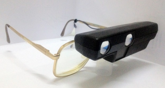
Our patient’s previous autofocus telescope and prescription spectacles.
The distance spectacle prescription for her right eye was −7.00/-3.25×170 with visual acuity of 6/48 and for the left eye was −8.00/-2.00×175 with visual acuity of 6/48. Near visual acuity for both right eye and left eye was N10 at 20cm. Subjective refraction was found to be similar to her spectacle prescription on presentation, and after conducting a pinhole test did not show any further improvement.
Low-vision assessment was carried out and the BITA® Vision Enhancer 3/8 4× binocularly was introduced and fitted to her distance spectacles (Figure 2 ). Our patient found that the BITA® telemicroscope was acceptable and her visual acuity (VA) for both eyes was 6/9 when using the BITA® device. The BITA® telemicroscope was light in weight compared to her auto-focus telescopes. It was also smaller in size and therefore not readily noticeable by others when in use. The BITA® telemicroscope was therefore prescribed to our patient.
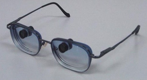
Bi-level telemicroscopic apparatus (BITA® ) telemicroscopes and prescription spectacles, newly prescribed to our patient.
A slit lamp examination showed that our patient could be fitted with contact lenses (CLs). Her tear prism was sufficient and the tear break-up time (TBUT) was 13 seconds for both eyes. In order to reduce glare, Flexcon prosthetic iris-tinted CM Brown color CLs 38% (12mm iris diameter with 3.5mm clear pupil size) /8.4/14.00/plano/ were prescribed to our patient for both eyes (Figure 3 ). No prescription was incorporated into the CLs because our patient wanted to use the CLs either with her distance spectacles or with the BITA® telemicroscope. Her vision when using the CLs and her own spectacles improved one line to 6/36 for both eyes. The CLs were prescribed with a multi-purpose solution as the disinfecting system. Our patient returned for aftercare following one week of CL usage. She reported that with the prosthetic CLs, glare was no longer a major issue, her nystagmus was reduced and her vision was clearer with her spectacles. She also reported that her vision was clearer when she used the iris-tinted CLs with the BITA® telemicroscope. Our patient was advised to return for routine CL aftercare every three months, as is advised to other CL wearers.
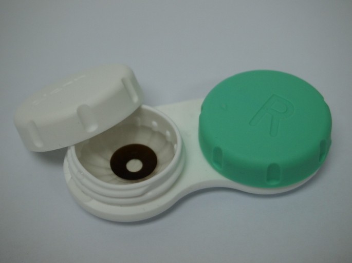
Prosthetic tinted contact lenses with clear pupil area used by our patient.
The issue of glare, sensitivity to light and occurrence of nystagmus in patients with albinism is the result of overstimulation of the hypopigmented retina under normal circumstances. The tinted iris pattern imprinted on the CL reduces the amount of light that enters the eye and thus alleviated our patient’s sensitivity to light and her problem with glare. The reduced light entering her eye, together with the 3.5mm clear pupil on the prescribed CL, forms a smaller blur circle on the fovea hence reducing the scanning that induces our patient’s nystagmus.
Patients with albinism generally have hypopigmentation of the retina, therefore under normal circumstances there is a relative increase in the absorption of light because of the reduced pigment in the retina. As a result, the hypopigmented retina is relatively more stimulated compared to a normally pigmented retina. Hence, a reduction in the light entering the eye and falling onto the hypopigmented retina would cause a subjective improvement in vision, as was described by our patient and demonstrated by her distance vision improving by one Snellen line. The reduced squinting in our patient was observed when she entered a brightly illuminated area while using the CLs. The reduced amount of light entering the eye caused by the use of the CLs delivered a positive benefit to the visual ability of our patient. Similar findings were observed by Philips in a severe albino patients fitted with soft prosthetic contact lens that reduced both the photophobic symptoms and enhanced facial cosmesis [ 9 ].
CL aftercare is very important and serves to ensure that the patient’s eyes are closely monitored. Since our patient has nystagmus, complications caused by CL use have to be expected. Frequent CL aftercare appointments are warranted in order to minimize any potential CL complications. Patient education on lens care and maintenance is also needed to ensure that their eyes stay healthy and to avoid any possible eye infection. Consequently, the need for continued CL aftercare was emphasized to our patient so that she could continue using the prosthetic CLs with a minimum of complications.
When the prosthetic CLs were used together with the BITA® telemicroscope, our patient reported that she had clearer vision. The clearer vision could be explained by the improvement in her binocular vision. It is interesting to note that in some cases of nystagmus, blurring of vision in one eye (instead of occluding the eye) will improve the vision of the other eye. The use of the telemicroscope itself does cause some pupillary constriction, thus reducing the light entering the eye and reducing the stimulation of the fovea, hence causing the subjective improvement in vision that our patient reported.
The BITA® telemicroscope is very small (smaller than a paper clip) and much lighter compared to our patient’s old auto-focus telescope. It was therefore not surprising that the BITA® telemicroscope was easily accepted by our patient. This made our patient more confident and more willing to accept using low-vision aids. Ultimately, this combination of management has enabled our patient to cope better with her outdoor activities and to do so in a more productive manner. A previous study has demonstrated that the BITA® telemicroscope is small in size and therefore not generally noticeable to an onlooker. The spectacle-mounted BITA® telemicroscopes allow patients to see a magnified view through the device while simultaneously seeing a normal view through the spectacles [ 10 ]. The BITA® telemicroscope is capable of remaining in focus for distances from about three feet to infinity without the need for any adjustment. It can also be adjusted for near work by turning the barrel of each scope to focus. As the telemicroscopes are very small and situated at the upper part of the lenses, they look almost like normal spectacles, as can be seen in Figure 2 . A special tint on the spectacle lenses can further camouflage the telescopic device so that it becomes less noticeable to onlookers.
Contact lenses with special iris tinting and a clear pupil area can be introduced to patients with albinism and can benefit these patients by reducing the amount of light entering the hypopigmented eye, thereby reducing the symptoms of glare and sensitivity to light. However, care and diligence is needed to assess the suitability and usefulness of the tinted contact lenses so that they fulfill a rehabilitative role in patients with albinism. The BITA® telemicroscopes fitted to the prescription spectacles appeared more cosmetically acceptable to our patient, and were able to facilitate our patient in coping with her daily activities thus improving her quality of life through clearer distance vision.
Written informed consent was obtained from the patient for publication of this case report and any accompanying images. A copy of the written consent is available for review by the Editor-in-Chief of this journal.
Oetting WS, Fryer JP, Shriram S, King RA: Oculocutaneous albinism type 1: The last 100 years. Pigment Cell Res. 2003, 16: 307-311. 10.1034/j.1600-0749.2003.00045.x.
Article CAS PubMed Google Scholar
Hagen EAH, Houston GC, Hoffmann MB, Jeffery G, Morland AB: Retinal abnormalities in human albinism translate into a reduction of grey matter in the occipital cortex. Eur J Neurosci. 2005, 22: 2475-2480. 10.1111/j.1460-9568.2005.04433.x.
Article Google Scholar
Hagen EAH, Houston GC, Hoffmann MB, Morland AB: Pigmentation predicts the shift in the line of decussation in humans with albinism. Eur J Neurosci. 2007, 25: 503-511. 10.1111/j.1460-9568.2007.05303.x.
Blachford SL A: The Gale Encyclopedia of Genetic Disorders. 2002, Gale Group-Thompson Learning, Detroit, MI
Google Scholar
Wildsoet CF, Oswald PJ, Clark S: Albinism: its implications for refractive development. Invest Ophthalmol Vis Sci. 2007, 41: 1-7.
Neveu MM, Holder GE, Sloper JJ, Jeffery G: Optic chiasm formation in humans is independent of foveal development. Eur J Neurosci. 2005, 22: 1825-1829. 10.1111/j.1460-9568.2005.04364.x.
Article PubMed Google Scholar
Omar R, Fatin NN N: Assessment of Quality of Life among Albinos (Penilaian Tahap Kualiti Hidup di kalangan Albino). [ http://www.ukm.academia.edu/RokiahOmar/Papers/247035/Penilaian_tahap_kualti_hidup_albino# ]
Sowka JW, Gurwood AS: Low vision rehabilitation of the albino patient. J Am Optom Assoc. 1991, 62: 533-536.
CAS PubMed Google Scholar
Philips AJ: A prosthetic contact lens in the treatment of ocular manifestations of albinism. Clin Exp Opt. 1989, 72: 32-34. 10.1111/j.1444-0938.1989.tb03856.x.
Peli E, Vargas-Martin F: In-the-spectacle-lens telescopic device. J Biomed Opt. 2008, 13: 034027-10.1117/1.2940360.
Article PubMed PubMed Central Google Scholar
Download references
Acknowledgments
BITA® Vision Enhancer 3/8” 4.00X, supplied by: Conforma Contact Lenses. Low Vision, 4705 Colley Avenue, Norfolk, VA 23508, United States of America, Tel: 18004261700. Flexcon prosthetic iris-tinted CM Brown color CLs 38%, supplied by: Oculus (M) Sdn. Bhd. No: 19 Jalan PJS 11/14, Sunway Technology Park, 46150 Petaling Jaya, Malaysia, Tel: +603-56359737.
Author information
Authors and affiliations.
Optometry and Visual Science Program, School of Healthcare Sciences, Faculty of Health Sciences, Universiti Kebangsaan Malaysia, Jalan Raja Muda Abdul Aziz, 50300, Kuala Lumpur, Malaysia
Rokiah Omar & Siti Salwa Idris
Chung Optometry Consultant, 2-G-45, Wisma Rampai, Jalan 34/26, Rampai Town Centre, Setapak, 53300, Kuala Lumpur, Malaysia
Chung Kah Meng
Faculty of Medicine and Defence Health, National Defence University of Malaysia, Sg Besi Camp, 57000, Kuala Lumpur, Malaysia
Victor Feizal Knight
You can also search for this author in PubMed Google Scholar
Corresponding author
Correspondence to Rokiah Omar .
Additional information
Competing interests.
The authors declare that they have no competing interests.
Authors’ contributions
RO examined our patient, analyzed and interpreted investigative data and wrote the manuscript. SSI examined and analyzed the investigative data. CKM performed a literature review and contributed to writing the manuscript. VFK made critical revisions and contributed to the manuscript writing. All authors read and approved the final manuscript.
Authors’ original submitted files for images
Below are the links to the authors’ original submitted files for images.
Authors’ original file for figure 1
Authors’ original file for figure 2, authors’ original file for figure 3, authors’ original file for figure 4, authors’ original file for figure 5, rights and permissions.
This article is published under license to BioMed Central Ltd. This is an Open Access article distributed under the terms of the Creative Commons Attribution License ( http://creativecommons.org/licenses/by/2.0 ), which permits unrestricted use, distribution, and reproduction in any medium, provided the original work is properly cited.
Reprints and permissions
About this article
Cite this article.
Omar, R., Idris, S.S., Meng, C.K. et al. Management of visual disturbances in albinism: a case report. J Med Case Reports 6 , 316 (2012). https://doi.org/10.1186/1752-1947-6-316
Download citation
Received : 17 January 2012
Accepted : 03 August 2012
Published : 19 September 2012
DOI : https://doi.org/10.1186/1752-1947-6-316
Share this article
Anyone you share the following link with will be able to read this content:
Sorry, a shareable link is not currently available for this article.
Provided by the Springer Nature SharedIt content-sharing initiative
- Low-vision rehabilitation
- Special contact lenses
- Telemicroscopes
Journal of Medical Case Reports
ISSN: 1752-1947
- Submission enquiries: Access here and click Contact Us
- General enquiries: [email protected]

- Create account
Low Vision and Vision Rehabilitation in Glaucoma
Glaucoma is an optic neuropathy characterized by elevated intraocular pressure leading to visual field loss. It is the leading cause of irreversible blindness globally [1] . Visual impairment experienced by glaucoma patients can result in challenges with activities of daily living (ADLs), increased morbidity, and consequent negative impacts on mental health. [2] [3] [4] [5] [6] [7] While disease progression is targeted with medicine and surgery, irreversible vision related disabilities can be addressed through low-vision services (LVS). [8] This article presents information on how to recognize and respond to glaucoma patients with low-vision needs.
- 1 Definition of Low Vision
- 2 Glaucoma and Low Vision Epidemiology
- 3 Vision Impairment and Disability
- 4.1 History
- 4.2 Visual Acuity
- 4.3 Near Acuities
- 4.4 Visual Field
- 4.5 Contrast Sensitivity
- 4.6 Observation of Patient
- 5.1 Near magnification [7]
- 5.2 Field Enhancement
- 5.3 Glare control
- 5.4 ADL Assistance
- 5.5 Mental Health Impairments
- 6 Treatment Outcomes
- 7 Additional Resources
- 8 References
Definition of Low Vision
According to the American Academy of Ophthalmology’s Preferred Practice Patterns (PPP) for Vision Rehabilitation, low vision is defined as a visual impairment caused by eye disease in which visual acuity is 20/50 or worse in the better-seeing eye and cannot be corrected or improved with regular eyeglasses, contact lenses, medicine, or surgery. [9] It is further qualified as uncorrectable vision loss that may be better than 20/50 acuity, but which involves loss of visual field, reduced contrast sensitivity, increased glare, or difficulty with daily activities.
The International Classification of Diseases, Ninth Revision (ICD-9) and the International Classification of Diseases, Tenth Revision, Clinical Modification (ICD-10 CM) define low vision as patients who fall within Category of Visual Impairment 1 or 2, which includes patients with visual acuity 20/70 or worse in the better-seeing eye, or with any loss in visual field that impacts ADLs.
Glaucoma and Low Vision Epidemiology
An understanding of the epidemiology of glaucoma patients with low vision is limited to prevalence figures estimated through combined meta-analyses and census data ranging from 2010 to 2015. It is also limited by the way in which terms are defined, with glaucoma definitions varying by visual field testing or intraocular pressure measurements, and low vision being defined through the limited scope of visual acuity, which is often not the primary complaint in glaucoma.
In 2013, the number of people (40-80 years of age) worldwide with glaucoma was estimated at 64.3 million, projected to include 76.0 million by 2020 and 111.8 million by 2040. [1] The most up-to-date estimates for the United States indicate that there are 2.7 million Americans with glaucoma3, (1.9% of the population). Low vision (when defined as visual acuity 20/50 or worse in the better-seeing eye) was last estimated in 2010 to affect 2.9 million people in the United States (2.04% of the population). [10]
From a few studies defining symptomatic complaints in glaucoma, there is some understanding of the overlap of low vision in glaucoma patients. For example, a small survey study found that 57% of glaucoma patients reported needing more light, 55% reported blurry vision, 46% reported glare, 36% reported noticeable visual field losses, and 30% reported contrast sensitivity losses. [11] Glare, visual field losses, and contrast sensitivity losses would directly qualify patients as having low vision as defined by the Academy’s PPP, and as such are visual disabilities targetable by LVS. There are a significant number of people affected by glaucoma, and, of those patients, many are symptomatic in ways that meet the criteria for low vision. A cross-sectional study describing demographic and clinical characteristics of a glaucoma patient population attending vision rehabilitation found that patients reported the greatest difficulty with reading (88%), writing (72%), adn mobility (67%). Most glaucoma patients attending low vision rehabilitation were functionally monocular, but not legally blind. [12]

Vision Impairment and Disability
The loss of peripheral vision as detected by visual field testing is the most commonly followed measurement of glaucoma progression. The severity, magnitude, and rates of change in binocular visual field sensitivity are significantly correlated with quality-of-life measures in glaucoma patients, especially in later disease. [3] However, patients can experience a decline in quality of life and increase in disability even in early stages of glaucoma, and these changes may not always correlate with clinical measures.
The Collaborative Initial Glaucoma Treatment Study (CIGTS), found that more than 25% of newly diagnosed glaucoma patients report blurred vision, difficulty adapting between light and dark, trouble seeing in the dark, and problems with bright lights, and that visual field testing was only modestly correlated with patient complaints. [4] These findings underscore the importance of screening glaucoma patients for visual disability outside of clinical testing. Vision related quality of life may be a more useful patient-centered outcome measurement, as well as something that can be targeted through LVS. [4]
The most common glaucoma patient complaints are difficulties with mobility, reading, and driving. [5] When glaucoma patients were surveyed about their mobility, a review found 49% of patients have difficulty with steps, 42% with shopping, and 36% with crossing the road. [6] This difficulty was correlated with a significant increase in falls, entry into assisted living, restricted physical activity, and a decreased quality of life, leading to an overall increase in morbidity and mortality. [6] For glaucoma patients surveyed about reading 40% of patients endorse general difficulty. [6] Research examining the extent and cause of the reading deficit found reading difficulty in glaucoma patients even with retained acuity, difficulty following a line or moving to the next line, difficulty with small print or low contrast, and decreased reading speeds to the point of reading impairment (less than 80 words per minute) in sustained silent reading. [13] [14] [15] Finally, glaucoma patients surveyed about driving reported increased perceived difficulty with driving, particularly at night, with mixed data on increased risk of collisions and resulting morbidity and mortality. However, glaucoma patients are three times more likely to stop driving due to their perceived deficits, which is associated with higher rates of entry into assisted living, lower quality of life, and depression. The resulting loss of function in all three categories of mobility, reading, and driving is correlated with increased rates of depression and anxiety. [5]
Evaluation of Low Vision in Glaucoma
Screen for patient functional complaints, patient quality of life, and ability to perform ADLs. [9]
- Activities of Daily Vision Scale (ADVS)
- National Eye Institute Visual Functioning Questionnaire (NEI-VFQ)
- Visual Function Index (VF-14)
- Visual Activities Questionnaire (VAQ)
- Glaucoma Quality of Life (GQL-15)
Visual Acuity
Test with high contrast charts and bright lighting. Encourage the patient to shift their gaze or move their head to find the optimum fixation point for best vision. [5]
- Projection charts: Not appropriate due to low contrast and presentation in a dark room
- Sloan chart: Use brightly lit at 10 ft
- Early Treatment Diabetic Retinopathy Study (ETDRS) chart: For visual acuity <20/100
- Designs for Vision Distance Test Chart for the Partially Sighted: For severe acuity loss
Near Acuities
Carefully evaluate due to common patient complaints regarding reading. [7]
- Bailey-Lovie Near Reading Card: Tests whole word reading acuity, more useful in evaluating patients for reading aids than single letter acuities
Visual Field
Evaluate what remaining field is available for rehabilitation. [7]
- Goldmann perimeter: Large stimuli for easier assessment
- Humphrey automated 10-2 field: For remaining central field in advanced glaucoma
Contrast Sensitivity
Important for resolving objects in daily visual tasks, such as recognizing a face, distinguishing between pills, or resolving where steps begin and end [7]
- Pelli-Robson Contrast Sensitivity Chart: One size letters with decreasing contrast, patients must be able to see 40M-sized letters
- VISTECH contrast test: Sine-wave/bar patterns for vision less than 20/40
Observation of Patient
Observe the patient performing visual tasks, e.g., reading, writing, walking, navigating steps. [5]
- Quality of continuous reading, reading speeds, print size, errors in certain field locations
- Speed of walking, head swing and room scanning, missed objects/bumping into items
Management of Glaucoma Patients with Low Vision
The goal of LVS is to increase the patient’s ability to maximize function with their remaining vision. [9]
Near magnification [7]
- Handheld/stand/electronic magnifiers: Electronic magnifiers are the gold standard in LVS due to variable magnification, high contrast, and black/white reversible polarity
- Computer Screen magnification programs, screen readers
Field Enhancement
- Scanning: Head swings, eye sweeps, slower approach speed. Helps patients to increase their awareness of the areas that need to be processed and develop a systematic plan for search patterns
- Field Expanders: Minifiers, reverse telescopes, or prisms to reduce the entire field into the central field or transfer the peripheral field onto the central field. Helps patients in static scenarios (spotting objects, viewing from a stable position), not for dynamic use (walking)
Glare control
- Discomfort glare: Causes discomfort and reduces visual task efficiency but not resolution
- Manage by turning down lights, adjusting angle of incoming light, absorptive lenses
- Disability glare: Reduces the resolution/ ability to identify visual stimulus
- Manage with polarizing lenses, anti-reflective coatings, incandescent lighting
ADL Assistance
- Referred retinal locus and eccentric fixation training for preserved, non-central vision
- Room lighting: Even, without shadow, adequate for mobility and ADLs but without glare or reflections
- For missing visual field: Typoscope, signature guide, place markers
- Tactile aids: Dots, plasticized marking pens, bold line markers
- Talking devices: Watches, clocks, calculators, phones
- Orientation and mobility training
Mental Health Impairments
- Networking, social support system, team concept: Important for combating increased rates of depression and anxiety
Treatment Outcomes
LVS treatment outcomes have been evaluated using clinical questionnaires in multiple studies prior to and after completion of 3 to 9 months of therapy. A wide range of measures have been used, and many categories were found to be improved by a statistically significant amount from baseline following treatment including overall visual ability, reading, visual information processing, mobility, near tasks, social functioning, and emotional well-being. [16] [17] [18] The improvements in overall visual ability in particular showed clinically meaningful improvements in nearly half of the patients attending LVS. [16] The magnitude of the improvement in all categories and across studies was characterized as moderate. [16] [17]
However, these data are for all patients utilizing LVS and the majority of LVS referrals are patients with central vision loss, such as in age-related macular degeneration (AMD). [19] In one study evaluating the effect of LVS on glaucoma patients, those with best-corrected visual acuity (BCVA) between 20/70 and 20/400 in the better-seeing eye and a diagnosis of glaucoma were randomized to either low-vision examination and treatment or only low-vision examination. [18] A significant improvement was found in reading ability and overall visual ability following LVS when compared to the control group. [18]
The data for low vision in glaucoma patients is still developing. It is clear that there is a large population of glaucoma patients, many of which have visual functional deficits that meet criteria for low vision. In the setting of these irreversible deficits, patient disability can be targeted through low vision examination and services. It is important to identify these patients, discuss strategies for improved functionality, and refer them to the appropriate LVS in a timely fashion.
Additional Resources
- Turbert D, and Dan Gudgel John D Shepherd, Mary Lou Jackson, Linda M Lawrence, Terry L Schwartz. Low Vision . American Academy of Ophthalmology. EyeSmart/Eye health. https://www.aao.org/eye-health/diseases/low-vision-list . Accessed March 14, 2019.
- ↑ 1.0 1.1 Tham, Yih-Chung, et al. Global prevalence of glaucoma and projections of glaucoma burden through 2040: a systematic review and meta-analysis. Ophthalmology 121.11 (2014): 2081-2090.
- ↑ Cesareo, Massimo, et al. Visual disability and quality of life in glaucoma patients. Progress in Brain Research 221 (2015): 359-374.
- ↑ 3.0 3.1 Medeiros, Felipe A., et al. Longitudinal changes in quality of life and rates of progressive visual field loss in glaucoma patients. Ophthalmology 122.2 (2015): 293-301.
- ↑ 4.0 4.1 4.2 Mills, Richard P., et al. Correlation of visual field with quality-of-life measures at diagnosis in the Collaborative Initial Glaucoma Treatment Study (CIGTS). Journal of Glaucoma 10.3 (2001): 192-198.
- ↑ 5.0 5.1 5.2 5.3 5.4 Pabon, Sheila, et al. Low Vision Therapy for Glaucoma Patients. Current Ophthalmology Reports (2017): 1-8.
- ↑ 6.0 6.1 6.2 6.3 Ramulu, Pradeep. Glaucoma and disability: which tasks are affected, and at what stage of disease? Current Opinion in Ophthalmology 20.2 (2009): 92.
- ↑ 7.0 7.1 7.2 7.3 7.4 Robison, Scott. Advanced Glaucoma and Low Vision: Evaluation and Treatment. The Glaucoma Book. Springer New York, 2010. 351-381.
- ↑ Renieri, Giulia, et al. Changes in quality of life in visually impaired patients after low-vision rehabilitation. International Journal of Rehabilitation Research 36.1 (2013): 48-55.
- ↑ 9.0 9.1 9.2 American Academy of Ophthalmology Vision Rehabilitation Committee. Preferred Practice Pattern Guidelines. Vision Rehabilitation. San Francisco, CA: American Academy of Ophthalmology; 2013.
- ↑ Massof, Robert W. A model of the prevalence and incidence of low vision and blindness among adults in the US. Optometry & Vision Science 79.1 (2002): 31-38.
- ↑ Hu, Cindy X., et al. What do patients with glaucoma see? Visual symptoms reported by patients with glaucoma. The American Journal of the Medical Sciences 348.5 (2014): 403-409.
- ↑ Kaleem MA, Rajjoub R, Schiefer C, et al. Characteristics of Glaucoma Patients Attending a Vision Rehabilitation Service. Ophthalmol Glaucoma. 2021;S2589-4196(21)00064-8.
- ↑ Viswanathan, Ananth C., et al. Severity and stability of glaucoma: patient perception compared with objective measurement. Archives of Ophthalmology 117.4 (1999): 450-454.
- ↑ Altangerel, Undraa, et al. Assessment of function related to vision (AFREV). Ophthalmic epidemiology 13.1 (2006): 67-80.
- ↑ Ramulu, Pradeep Y., et al. Difficulty with out-loud and silent reading in glaucoma. Investigative ophthalmology & visual science 54.1 (2013): 666-672.
- ↑ 16.0 16.1 16.2 Goldstein, Judith E., et al. Clinically meaningful rehabilitation outcomes of low vision patients served by outpatient clinical centers. JAMA Ophthalmology 133.7 (2015): 762-769.
- ↑ 17.0 17.1 Lamoureux, Ecosse L., et al. The effectiveness of low-vision rehabilitation on participation in daily living and quality of life. Investigative Ophthalmology & Visual Science 48.4 (2007): 1476-1482.
- ↑ 18.0 18.1 18.2 Patodia, Yogesh, et al. Clinical effectiveness of currently available low-vision devices in glaucoma patients with moderate-to-severe vision loss. Clinical Ophthalmology 11 (2017): 683.
- ↑ Kaleem, Mona A., et al. Referral to Low Vision Services for Glaucoma Patients: Referral Patterns and Characteristics of Those Who Refer. Journal of Glaucoma (2017).
Low-vision intervention in individuals with age-related macular degeneration
Affiliations.
- 1 Shanmugha Arts, Science, Technology and Research Academy (SASTRA) University, Thanjavur; Department of Optometry, Low Vision Care Clinic, Sankara Nethralaya, Chennai, Tamil Nadu, India.
- 2 Shri Bhagwan Mahavir Vitreoretinal Services, Sankara Nethralaya, Chennai, Tamil Nadu, India.
- PMID: 32317472
- PMCID: PMC7350438
- DOI: 10.4103/ijo.IJO_1093_19
Purpose: The objective of this study was to estimate the level of visual impairment in patients diagnosed to have age-related macular degeneration (ARMD) who presented to low-vision care (LVC) clinic at a tertiary eye care center in India, to analyze the type of distant and near devices prescribed to them and to compare the visual benefit in different age groups among patients with ARMD.
Methods: A retrospective review was done for 91 patients with low-vision secondary to ARMD who were referred to the LVC clinic from 2016 to 2017. Demographic profile: age, gender, occupation, ocular history, visual acuity status, and type of low-vision device (LVD) preferred were documented. The details of LVDs and subsequent improvements were noted.
Result: Of the 91 patients, 64 (70.3%) were men and 27 (29.7%) were women. Of the cases which were referred, 36.26% had a severe visual impairment (VI), 32.96% had moderate VI, 28.57% had mild VI, and 5.49% had profound VI. The majority of the patients had myopia 57 (62.63%), followed by hyperopia in 25 (27.47%) subjects. The subjects were divided into three groups based on age 40-65 years, 66-75 years, and above 75 years for the analysis of VI. There was a statistically significant improvement (P < 0.01) in near vision with the help of LVDs in all three groups. SEE TV binocular telescope was the most commonly prescribed LVD for viewing distant objects. The most commonly preferred magnifier for near work was half-eye spectacle (56%) followed by stand magnifier (9.9%) and portable video magnifier (9.9%).
Conclusion: The use of LVDs can help these patients with ARMD in cases where medical and surgical treatment have no or a limited role in restoring useful vision.
Keywords: Age-related macular degeneration; dome magnifier; low-vision; spectacle magnifier.
- India / epidemiology
- Macular Degeneration* / complications
- Macular Degeneration* / diagnosis
- Macular Degeneration* / epidemiology
- Middle Aged
- Retrospective Studies
- Vision, Low* / epidemiology
- Vision, Low* / etiology
- Visual Acuity
A generative AI reset: Rewiring to turn potential into value in 2024
It’s time for a generative AI (gen AI) reset. The initial enthusiasm and flurry of activity in 2023 is giving way to second thoughts and recalibrations as companies realize that capturing gen AI’s enormous potential value is harder than expected .
With 2024 shaping up to be the year for gen AI to prove its value, companies should keep in mind the hard lessons learned with digital and AI transformations: competitive advantage comes from building organizational and technological capabilities to broadly innovate, deploy, and improve solutions at scale—in effect, rewiring the business for distributed digital and AI innovation.
About QuantumBlack, AI by McKinsey
QuantumBlack, McKinsey’s AI arm, helps companies transform using the power of technology, technical expertise, and industry experts. With thousands of practitioners at QuantumBlack (data engineers, data scientists, product managers, designers, and software engineers) and McKinsey (industry and domain experts), we are working to solve the world’s most important AI challenges. QuantumBlack Labs is our center of technology development and client innovation, which has been driving cutting-edge advancements and developments in AI through locations across the globe.
Companies looking to score early wins with gen AI should move quickly. But those hoping that gen AI offers a shortcut past the tough—and necessary—organizational surgery are likely to meet with disappointing results. Launching pilots is (relatively) easy; getting pilots to scale and create meaningful value is hard because they require a broad set of changes to the way work actually gets done.
Let’s briefly look at what this has meant for one Pacific region telecommunications company. The company hired a chief data and AI officer with a mandate to “enable the organization to create value with data and AI.” The chief data and AI officer worked with the business to develop the strategic vision and implement the road map for the use cases. After a scan of domains (that is, customer journeys or functions) and use case opportunities across the enterprise, leadership prioritized the home-servicing/maintenance domain to pilot and then scale as part of a larger sequencing of initiatives. They targeted, in particular, the development of a gen AI tool to help dispatchers and service operators better predict the types of calls and parts needed when servicing homes.
Leadership put in place cross-functional product teams with shared objectives and incentives to build the gen AI tool. As part of an effort to upskill the entire enterprise to better work with data and gen AI tools, they also set up a data and AI academy, which the dispatchers and service operators enrolled in as part of their training. To provide the technology and data underpinnings for gen AI, the chief data and AI officer also selected a large language model (LLM) and cloud provider that could meet the needs of the domain as well as serve other parts of the enterprise. The chief data and AI officer also oversaw the implementation of a data architecture so that the clean and reliable data (including service histories and inventory databases) needed to build the gen AI tool could be delivered quickly and responsibly.

Creating value beyond the hype
Let’s deliver on the promise of technology from strategy to scale.
Our book Rewired: The McKinsey Guide to Outcompeting in the Age of Digital and AI (Wiley, June 2023) provides a detailed manual on the six capabilities needed to deliver the kind of broad change that harnesses digital and AI technology. In this article, we will explore how to extend each of those capabilities to implement a successful gen AI program at scale. While recognizing that these are still early days and that there is much more to learn, our experience has shown that breaking open the gen AI opportunity requires companies to rewire how they work in the following ways.
Figure out where gen AI copilots can give you a real competitive advantage
The broad excitement around gen AI and its relative ease of use has led to a burst of experimentation across organizations. Most of these initiatives, however, won’t generate a competitive advantage. One bank, for example, bought tens of thousands of GitHub Copilot licenses, but since it didn’t have a clear sense of how to work with the technology, progress was slow. Another unfocused effort we often see is when companies move to incorporate gen AI into their customer service capabilities. Customer service is a commodity capability, not part of the core business, for most companies. While gen AI might help with productivity in such cases, it won’t create a competitive advantage.
To create competitive advantage, companies should first understand the difference between being a “taker” (a user of available tools, often via APIs and subscription services), a “shaper” (an integrator of available models with proprietary data), and a “maker” (a builder of LLMs). For now, the maker approach is too expensive for most companies, so the sweet spot for businesses is implementing a taker model for productivity improvements while building shaper applications for competitive advantage.
Much of gen AI’s near-term value is closely tied to its ability to help people do their current jobs better. In this way, gen AI tools act as copilots that work side by side with an employee, creating an initial block of code that a developer can adapt, for example, or drafting a requisition order for a new part that a maintenance worker in the field can review and submit (see sidebar “Copilot examples across three generative AI archetypes”). This means companies should be focusing on where copilot technology can have the biggest impact on their priority programs.
Copilot examples across three generative AI archetypes
- “Taker” copilots help real estate customers sift through property options and find the most promising one, write code for a developer, and summarize investor transcripts.
- “Shaper” copilots provide recommendations to sales reps for upselling customers by connecting generative AI tools to customer relationship management systems, financial systems, and customer behavior histories; create virtual assistants to personalize treatments for patients; and recommend solutions for maintenance workers based on historical data.
- “Maker” copilots are foundation models that lab scientists at pharmaceutical companies can use to find and test new and better drugs more quickly.
Some industrial companies, for example, have identified maintenance as a critical domain for their business. Reviewing maintenance reports and spending time with workers on the front lines can help determine where a gen AI copilot could make a big difference, such as in identifying issues with equipment failures quickly and early on. A gen AI copilot can also help identify root causes of truck breakdowns and recommend resolutions much more quickly than usual, as well as act as an ongoing source for best practices or standard operating procedures.
The challenge with copilots is figuring out how to generate revenue from increased productivity. In the case of customer service centers, for example, companies can stop recruiting new agents and use attrition to potentially achieve real financial gains. Defining the plans for how to generate revenue from the increased productivity up front, therefore, is crucial to capturing the value.
Upskill the talent you have but be clear about the gen-AI-specific skills you need
By now, most companies have a decent understanding of the technical gen AI skills they need, such as model fine-tuning, vector database administration, prompt engineering, and context engineering. In many cases, these are skills that you can train your existing workforce to develop. Those with existing AI and machine learning (ML) capabilities have a strong head start. Data engineers, for example, can learn multimodal processing and vector database management, MLOps (ML operations) engineers can extend their skills to LLMOps (LLM operations), and data scientists can develop prompt engineering, bias detection, and fine-tuning skills.
A sample of new generative AI skills needed
The following are examples of new skills needed for the successful deployment of generative AI tools:
- data scientist:
- prompt engineering
- in-context learning
- bias detection
- pattern identification
- reinforcement learning from human feedback
- hyperparameter/large language model fine-tuning; transfer learning
- data engineer:
- data wrangling and data warehousing
- data pipeline construction
- multimodal processing
- vector database management
The learning process can take two to three months to get to a decent level of competence because of the complexities in learning what various LLMs can and can’t do and how best to use them. The coders need to gain experience building software, testing, and validating answers, for example. It took one financial-services company three months to train its best data scientists to a high level of competence. While courses and documentation are available—many LLM providers have boot camps for developers—we have found that the most effective way to build capabilities at scale is through apprenticeship, training people to then train others, and building communities of practitioners. Rotating experts through teams to train others, scheduling regular sessions for people to share learnings, and hosting biweekly documentation review sessions are practices that have proven successful in building communities of practitioners (see sidebar “A sample of new generative AI skills needed”).
It’s important to bear in mind that successful gen AI skills are about more than coding proficiency. Our experience in developing our own gen AI platform, Lilli , showed us that the best gen AI technical talent has design skills to uncover where to focus solutions, contextual understanding to ensure the most relevant and high-quality answers are generated, collaboration skills to work well with knowledge experts (to test and validate answers and develop an appropriate curation approach), strong forensic skills to figure out causes of breakdowns (is the issue the data, the interpretation of the user’s intent, the quality of metadata on embeddings, or something else?), and anticipation skills to conceive of and plan for possible outcomes and to put the right kind of tracking into their code. A pure coder who doesn’t intrinsically have these skills may not be as useful a team member.
While current upskilling is largely based on a “learn on the job” approach, we see a rapid market emerging for people who have learned these skills over the past year. That skill growth is moving quickly. GitHub reported that developers were working on gen AI projects “in big numbers,” and that 65,000 public gen AI projects were created on its platform in 2023—a jump of almost 250 percent over the previous year. If your company is just starting its gen AI journey, you could consider hiring two or three senior engineers who have built a gen AI shaper product for their companies. This could greatly accelerate your efforts.
Form a centralized team to establish standards that enable responsible scaling
To ensure that all parts of the business can scale gen AI capabilities, centralizing competencies is a natural first move. The critical focus for this central team will be to develop and put in place protocols and standards to support scale, ensuring that teams can access models while also minimizing risk and containing costs. The team’s work could include, for example, procuring models and prescribing ways to access them, developing standards for data readiness, setting up approved prompt libraries, and allocating resources.
While developing Lilli, our team had its mind on scale when it created an open plug-in architecture and setting standards for how APIs should function and be built. They developed standardized tooling and infrastructure where teams could securely experiment and access a GPT LLM , a gateway with preapproved APIs that teams could access, and a self-serve developer portal. Our goal is that this approach, over time, can help shift “Lilli as a product” (that a handful of teams use to build specific solutions) to “Lilli as a platform” (that teams across the enterprise can access to build other products).
For teams developing gen AI solutions, squad composition will be similar to AI teams but with data engineers and data scientists with gen AI experience and more contributors from risk management, compliance, and legal functions. The general idea of staffing squads with resources that are federated from the different expertise areas will not change, but the skill composition of a gen-AI-intensive squad will.
Set up the technology architecture to scale
Building a gen AI model is often relatively straightforward, but making it fully operational at scale is a different matter entirely. We’ve seen engineers build a basic chatbot in a week, but releasing a stable, accurate, and compliant version that scales can take four months. That’s why, our experience shows, the actual model costs may be less than 10 to 15 percent of the total costs of the solution.
Building for scale doesn’t mean building a new technology architecture. But it does mean focusing on a few core decisions that simplify and speed up processes without breaking the bank. Three such decisions stand out:
- Focus on reusing your technology. Reusing code can increase the development speed of gen AI use cases by 30 to 50 percent. One good approach is simply creating a source for approved tools, code, and components. A financial-services company, for example, created a library of production-grade tools, which had been approved by both the security and legal teams, and made them available in a library for teams to use. More important is taking the time to identify and build those capabilities that are common across the most priority use cases. The same financial-services company, for example, identified three components that could be reused for more than 100 identified use cases. By building those first, they were able to generate a significant portion of the code base for all the identified use cases—essentially giving every application a big head start.
- Focus the architecture on enabling efficient connections between gen AI models and internal systems. For gen AI models to work effectively in the shaper archetype, they need access to a business’s data and applications. Advances in integration and orchestration frameworks have significantly reduced the effort required to make those connections. But laying out what those integrations are and how to enable them is critical to ensure these models work efficiently and to avoid the complexity that creates technical debt (the “tax” a company pays in terms of time and resources needed to redress existing technology issues). Chief information officers and chief technology officers can define reference architectures and integration standards for their organizations. Key elements should include a model hub, which contains trained and approved models that can be provisioned on demand; standard APIs that act as bridges connecting gen AI models to applications or data; and context management and caching, which speed up processing by providing models with relevant information from enterprise data sources.
- Build up your testing and quality assurance capabilities. Our own experience building Lilli taught us to prioritize testing over development. Our team invested in not only developing testing protocols for each stage of development but also aligning the entire team so that, for example, it was clear who specifically needed to sign off on each stage of the process. This slowed down initial development but sped up the overall delivery pace and quality by cutting back on errors and the time needed to fix mistakes.
Ensure data quality and focus on unstructured data to fuel your models
The ability of a business to generate and scale value from gen AI models will depend on how well it takes advantage of its own data. As with technology, targeted upgrades to existing data architecture are needed to maximize the future strategic benefits of gen AI:
- Be targeted in ramping up your data quality and data augmentation efforts. While data quality has always been an important issue, the scale and scope of data that gen AI models can use—especially unstructured data—has made this issue much more consequential. For this reason, it’s critical to get the data foundations right, from clarifying decision rights to defining clear data processes to establishing taxonomies so models can access the data they need. The companies that do this well tie their data quality and augmentation efforts to the specific AI/gen AI application and use case—you don’t need this data foundation to extend to every corner of the enterprise. This could mean, for example, developing a new data repository for all equipment specifications and reported issues to better support maintenance copilot applications.
- Understand what value is locked into your unstructured data. Most organizations have traditionally focused their data efforts on structured data (values that can be organized in tables, such as prices and features). But the real value from LLMs comes from their ability to work with unstructured data (for example, PowerPoint slides, videos, and text). Companies can map out which unstructured data sources are most valuable and establish metadata tagging standards so models can process the data and teams can find what they need (tagging is particularly important to help companies remove data from models as well, if necessary). Be creative in thinking about data opportunities. Some companies, for example, are interviewing senior employees as they retire and feeding that captured institutional knowledge into an LLM to help improve their copilot performance.
- Optimize to lower costs at scale. There is often as much as a tenfold difference between what companies pay for data and what they could be paying if they optimized their data infrastructure and underlying costs. This issue often stems from companies scaling their proofs of concept without optimizing their data approach. Two costs generally stand out. One is storage costs arising from companies uploading terabytes of data into the cloud and wanting that data available 24/7. In practice, companies rarely need more than 10 percent of their data to have that level of availability, and accessing the rest over a 24- or 48-hour period is a much cheaper option. The other costs relate to computation with models that require on-call access to thousands of processors to run. This is especially the case when companies are building their own models (the maker archetype) but also when they are using pretrained models and running them with their own data and use cases (the shaper archetype). Companies could take a close look at how they can optimize computation costs on cloud platforms—for instance, putting some models in a queue to run when processors aren’t being used (such as when Americans go to bed and consumption of computing services like Netflix decreases) is a much cheaper option.
Build trust and reusability to drive adoption and scale
Because many people have concerns about gen AI, the bar on explaining how these tools work is much higher than for most solutions. People who use the tools want to know how they work, not just what they do. So it’s important to invest extra time and money to build trust by ensuring model accuracy and making it easy to check answers.
One insurance company, for example, created a gen AI tool to help manage claims. As part of the tool, it listed all the guardrails that had been put in place, and for each answer provided a link to the sentence or page of the relevant policy documents. The company also used an LLM to generate many variations of the same question to ensure answer consistency. These steps, among others, were critical to helping end users build trust in the tool.
Part of the training for maintenance teams using a gen AI tool should be to help them understand the limitations of models and how best to get the right answers. That includes teaching workers strategies to get to the best answer as fast as possible by starting with broad questions then narrowing them down. This provides the model with more context, and it also helps remove any bias of the people who might think they know the answer already. Having model interfaces that look and feel the same as existing tools also helps users feel less pressured to learn something new each time a new application is introduced.
Getting to scale means that businesses will need to stop building one-off solutions that are hard to use for other similar use cases. One global energy and materials company, for example, has established ease of reuse as a key requirement for all gen AI models, and has found in early iterations that 50 to 60 percent of its components can be reused. This means setting standards for developing gen AI assets (for example, prompts and context) that can be easily reused for other cases.
While many of the risk issues relating to gen AI are evolutions of discussions that were already brewing—for instance, data privacy, security, bias risk, job displacement, and intellectual property protection—gen AI has greatly expanded that risk landscape. Just 21 percent of companies reporting AI adoption say they have established policies governing employees’ use of gen AI technologies.
Similarly, a set of tests for AI/gen AI solutions should be established to demonstrate that data privacy, debiasing, and intellectual property protection are respected. Some organizations, in fact, are proposing to release models accompanied with documentation that details their performance characteristics. Documenting your decisions and rationales can be particularly helpful in conversations with regulators.
In some ways, this article is premature—so much is changing that we’ll likely have a profoundly different understanding of gen AI and its capabilities in a year’s time. But the core truths of finding value and driving change will still apply. How well companies have learned those lessons may largely determine how successful they’ll be in capturing that value.

The authors wish to thank Michael Chui, Juan Couto, Ben Ellencweig, Josh Gartner, Bryce Hall, Holger Harreis, Phil Hudelson, Suzana Iacob, Sid Kamath, Neerav Kingsland, Kitti Lakner, Robert Levin, Matej Macak, Lapo Mori, Alex Peluffo, Aldo Rosales, Erik Roth, Abdul Wahab Shaikh, and Stephen Xu for their contributions to this article.
This article was edited by Barr Seitz, an editorial director in the New York office.
Explore a career with us
Related articles.

The economic potential of generative AI: The next productivity frontier

Rewired to outcompete

Meet Lilli, our generative AI tool that’s a researcher, a time saver, and an inspiration

IMAGES
VIDEO
COMMENTS
English: Living with Low Vision Powerpoint (VND.MS-POWERPOINT 5.51 MB) Spanish: Baja Vision PPT (VND.OPENXMLFORMATS-OFFICEDOCUMENT.PRESENTATIONML.PRESENTATION 5.22 MB) Use this 27-slide PowerPoint presentation to teach people about low vision, including how it's diagnosed and how people can adapt to low vision and maintain their independence.
A case study highlights the tertiary prevention (low vision examination and management) in a 5 year old boy with ROP related blindness to optimize his remaining visual capacity by using optical ...
Q4: Explain the use of electronic low vision aids as part of a management plan for patients with restricted visual fields and reduced visual acuity. One of the exciting developments in low vision is the use of new technology to provide access to information. Some involves the use of existing mainstream technology and others use purpose-built ...
Module 3: Low Vision. This module explains what low vision is, describes how people can find treatment and rehabilitation services, and offers tips for talking with eye care professionals about low vision. A slide presentation that provides an overview of low vision, including causes, symptoms, and treatment.
In this case, I was able to solve her problem easily and at very little cost to the patient, as she already had the solution in hand. William C., age 71, had a history of never doing well with low vision telescope glasses. He told me that when he wore a telescope it just made things larger, but equally blurry.
Free Google Slides theme and PowerPoint template. The design of this template is truly eye-catching! These creative slides are the perfect resource for innovative doctors or eye specialists who want to present ophthalmic clinical cases in a modern, fun way. This design includes lots of resources for health professionals so that you can present ...
The International Academy of Low Vision Specialists (IALVS) believes in life after vision loss.. We are a group of optometrists who are specially trained in low vision to help patients suffering from: Macular Degeneration: Wet or Dry, Albinism, Glaucoma, Stargardt's Disease, Diabetic Retinopathy, Retinitis Pigmentosa, and other Vision-Limiting Conditions
Decide which tasks are achievable based on the assessment of the patients ocular tissue. 5. Choose the appropriate low-vision device or series of devices. There are no hard-and-fast rules here. I always seek to design some form of optical system that provides patients with the greatest amount of latitude and function. 6.
Case presentation 3: RETINITIS PIGMENTOSA Mr. abc, 48 year old male was referred to the Low vision clinic by retinal clinic for low vision evaluation and management. He was diagnosed to have OU: Retinitis Pigmentosa. He was accompanied by his friend. By occupation he was sales person. Gross observation revealed he was not able to maintain eye ...
A low vision assessment determines the extent of vision loss and potential for vision rehabilitation. The specialist in low vision will assess the following: Your general health and eye health history. Functions of daily living related to your vision. Your visual acuity and other eye functions.
Retinitis Pigmentosa is a progressive condition resulting in a loss of vision. It may be associated with a number of systemic conditions or syndromes, or may be primary in nature. The rod-cone presentation is the more classic with a loss of peripheral vision to a resultant ring scotoma or tunnel vision, and a decrease in night vision, nyctalopia.
Title: Low Vision Reading and Comprehension: myReader Case Studies 1 Low Vision Reading and Comprehension myReader Case Studies. HumanWare ; Americas, Australasia, EMEA; 2 Presentation Overview. Limitations of CCTVs
Introduction A number of vision defects have been reported in association with albinism, such as photophobia, nystagmus and astigmatism. In many cases only prescription sunglasses are prescribed. In this report, the effectiveness of low-vision rehabilitation in albinism, which included prescription of multiple visual aids, is discussed. Case presentation We present the case of a 21-year-old ...
Low vision (when defined as visual acuity 20/50 or worse in the better-seeing eye) was last estimated in 2010 to affect 2.9 million people in the United States (2.04% of the population). From a few studies defining symptomatic complaints in glaucoma, there is some understanding of the overlap of low vision in glaucoma patients.
vision (i.e., low-vision individuals) have been largely unaddressed. To bridge this gap, we conducted a qualitative study that provides insights into how low-vision individuals experience visualizations. We found that participants utilized various strategies to examine visualizations using the screen magnifiersand also observed that
Purpose: The objective of this study was to estimate the level of visual impairment in patients diagnosed to have age-related macular degeneration (ARMD) who presented to low-vision care (LVC) clinic at a tertiary eye care center in India, to analyze the type of distant and near devices prescribed to them and to compare the visual benefit in different age groups among patients with ARMD.
Case presentation. A 21-year-old Malay woman diagnosed as having albinism presented to a Low Vision Clinic for low-vision assessment. Her problems were blurring of distant vision, glare, and her dissatisfaction with her current auto-focus spectacle-mounted telescope, which she reported as being heavy as well as cosmetically unacceptable (Figure ...
Central retinal vein occlusion (CRVO) is a significant cause of vision impairment and can occur at any age [ 1 ]. However, 90% of the CRVO patients are older than 50 years at the disease occurrence and only 10% of them are younger than 40 years [ 2 ]. The etiology can be quite varied, but age can be helpful in determining the differential ...
It's time for a generative AI (gen AI) reset. The initial enthusiasm and flurry of activity in 2023 is giving way to second thoughts and recalibrations as companies realize that capturing gen AI's enormous potential value is harder than expected.. With 2024 shaping up to be the year for gen AI to prove its value, companies should keep in mind the hard lessons learned with digital and AI ...